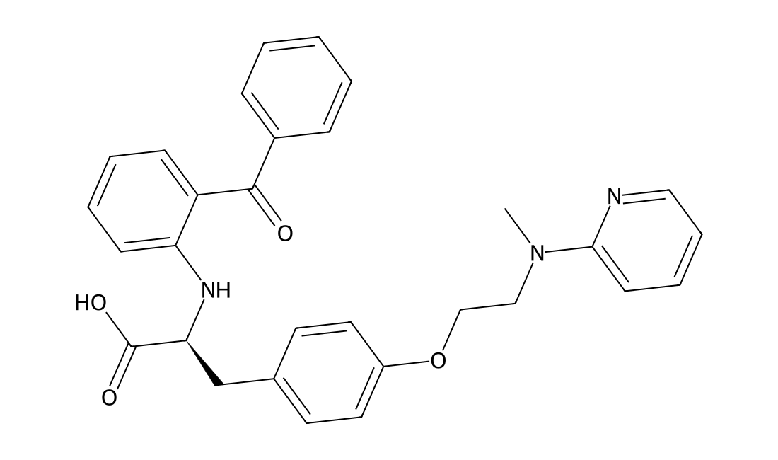
GW1929
CAS 196808-24-9
GW1929 is a potent PPAR-γ activator, with pKis of 8.84, < 5.5, and < 6.5 for human PPAR-γ, PPAR-α, and PPAR-δ, and pEC50s of 8.56 and 8.27 for human PPAR-γ and murine PPAR-γ, respectively.

Metabolic enzymes, lipid and glucose metabolism and hormone signaling.
Products may appear in more than one category. Counts refer to distinct catalog numbers within each category.
Other Research Tools (75) · Additional reagents, building blocks and specialized research tools.
Combine a keyword, research category and target to narrow the product list.
Loading catalog filters…
CAS 196808-24-9
GW1929 is a potent PPAR-γ activator, with pKis of 8.84, < 5.5, and < 6.5 for human PPAR-γ, PPAR-α, and PPAR-δ, and pEC50s of 8.56 and 8.27 for human PPAR-γ and murine PPAR-γ, respectively.
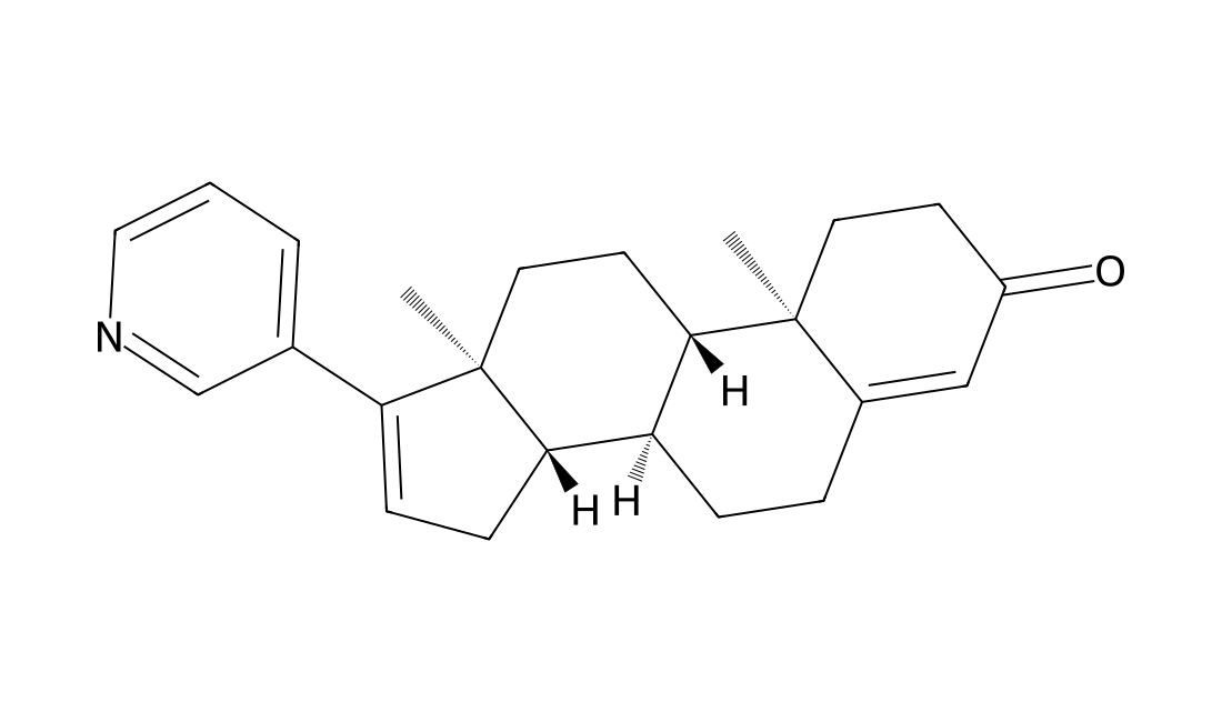
CAS 154229-21-7
D4-abiraterone is a major metabolite of abiraterone. D4-abiraterone is an inhibitor of CYP17A1, 3b-hydroxysteroid dehydrogenase (3βHSD) and steroid-5a-reductase (SRD5A) and also an antagonist of androgen receptor.
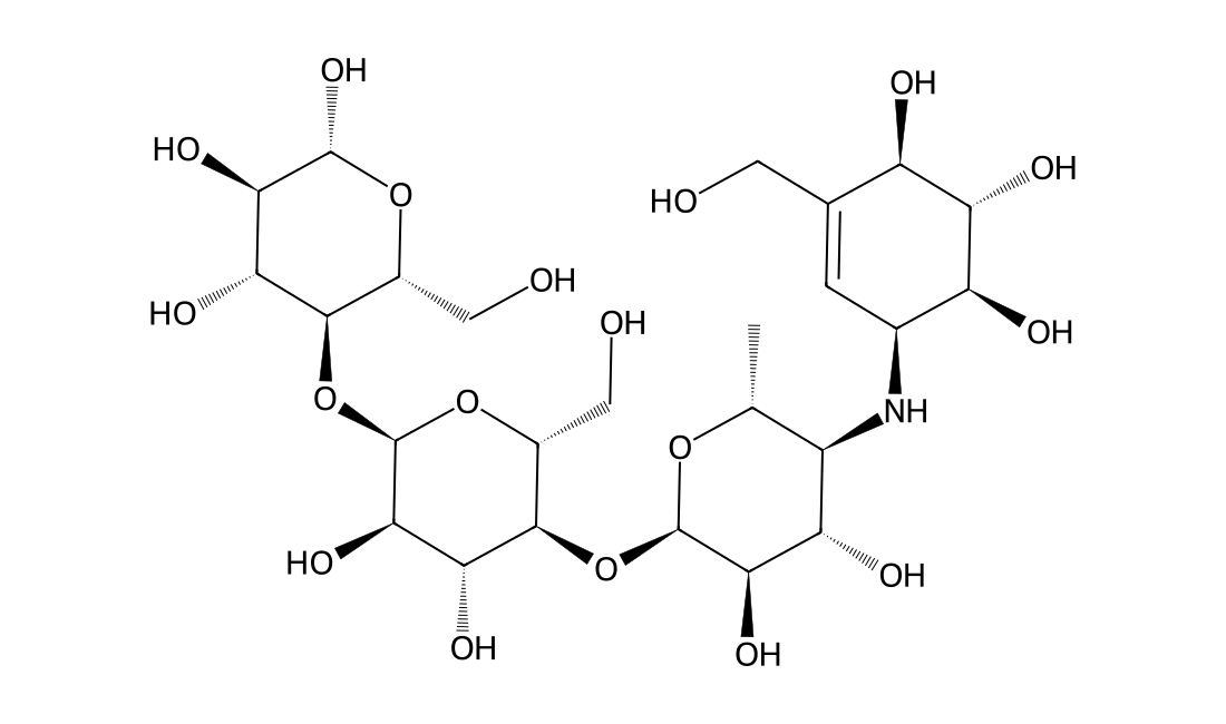
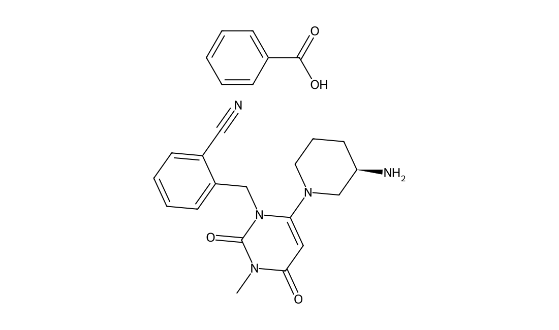
CAS 850649-62-6
Alogliptin benzoate(SYR 322) is a potent, selective inhibitor of DPP-4 with IC50 of <10 nM, exhibits greater than 10,000-fold selectivity over DPP-8 and DPP-9.
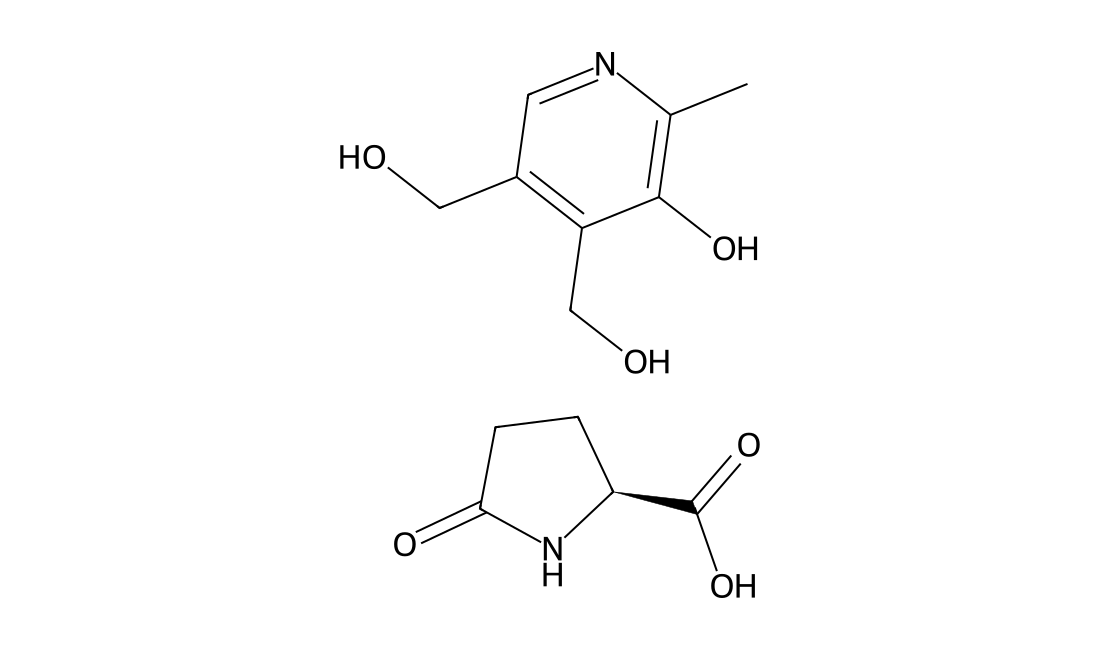
CAS 74536-44-0
In Vitro Simultaneous Metadoxine treatment notably inhibits adipogenic differentiation in a dose-dependent manner. Metadoxine treatment from day 4 through day 8 is required for inhibition of MDI-induced preadipocyte differentiation. Metadoxine prevents glutathione depletion and the increase in lipid peroxidation damage caused by ethanol and acetaldehyde in HepG2 cells. In hepatic stellate cells, Metadoxine prevents the increase in collagen and attenuated TNF-α secretion caused by acetaldehyde. Thus, Metadoxine could be useful in preventing the damage produced in early stages of alcoholic liver disease as it prevents the redox imbalance of the hepatocytes and prevents TNF-α induction, one of the earliest events in hepatic damage.
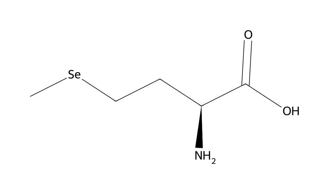
CAS 3211-76-5
L-SelenoMethionine is a major natural food-form of selenium.The median lethal dose (LD50) of L-SelenoMethionine in rats given an intraperitoneal injection was determined to 4.25 mg Se/kg body and thus is comparable to that of selenite or selenate. In mice, the LD50 of L-SelenoMethionine was 8.8 ± 1.37 mg Se/kg, and the minimal lethal dose, 4.0 mg Se/kg, after intravenous injection.
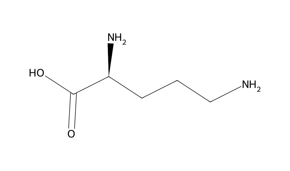
CAS 70-26-8
L-ornithine has an antifatigue effect by increasing the efficiency of energy consumption and promoting the excretion of ammonia. It is one of the key reactants in the urea cycle.
CAS 142-47-2
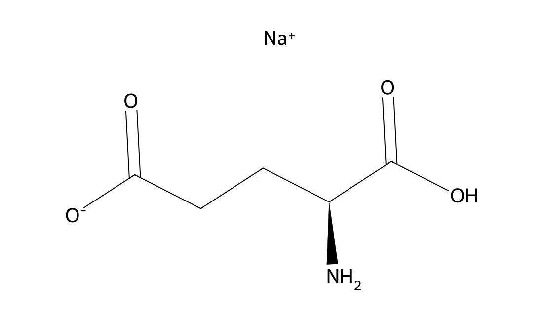
L-Glutamic acid monosodium salt is the sodium salt of glutamic acid, found naturally in tomatoes, cheese and other foods.
CAS 6133-30-8
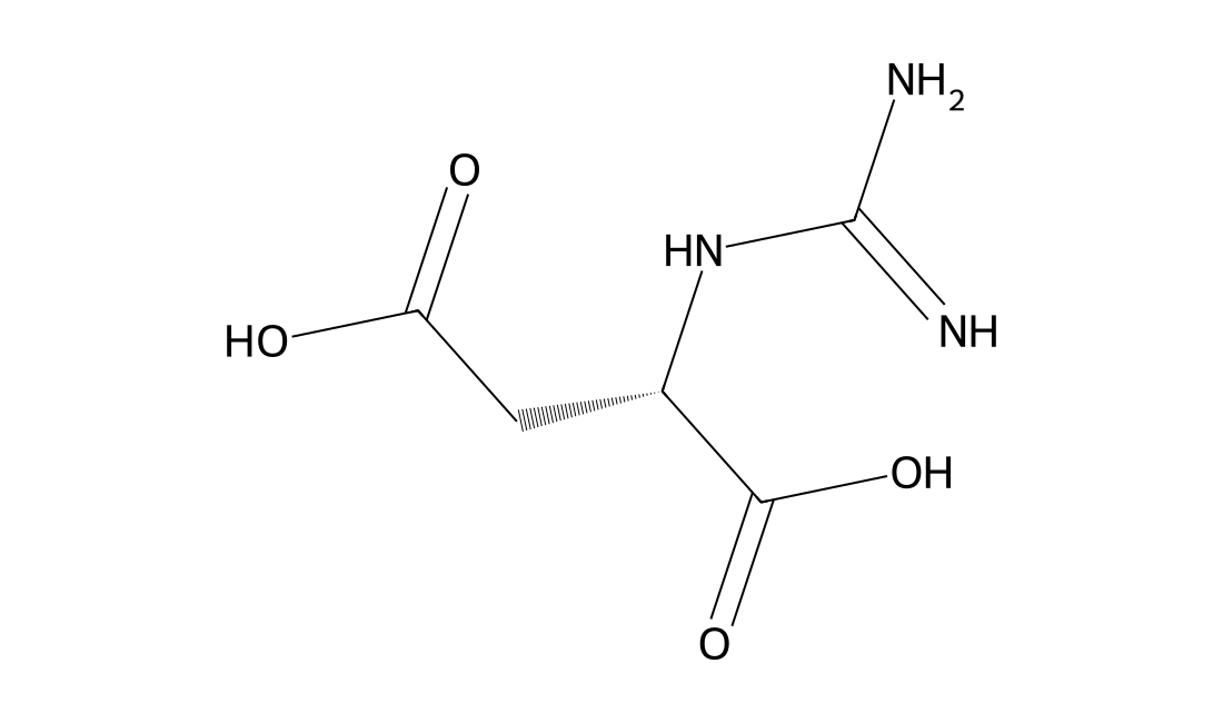
In Vivo Guanidinosuccinic acid, a constituent of normal urine, is elevated in the urine and serum of azotemic patients. Guanidinosuccinic acid (GSA), a guanidino compound found to be greatly increased in uremia, is administered by intraperitoneal (i.p.) injection to adult albino mice and to young mice 7, 14 and 21 days old. Epileptogenic and toxic properties are assessed and Guanidinosuccinic acid brain levels following i.p. injection ae determined. In adult mice, Guanidinosuccinic acid induces long-lasting generalized clonic and clonic-tonic convulsions in a dose-dependent manner with a CD50 (and 95% confidence interval) of 363 (287-458) mg/kg (n=35), and an LD50 of 579 (445-756) mg/kg. The CD50 of Guanidinosuccinic acid corresponded with a brain concentration of 56 nmol/g tissue. Electrocorticographic recording in five adult mice revealed epileptiform discharges (spikes, spike-waves, and polyspike-waves) which appeared concomitant with the convulsions, When young mice are i.p. injected with a (for adults) subconvulsive dose of Guanidinosuccinic acid (250 mg/kg), an age-dependent decrease is noted in Guanidinosuccinic acid-induced convulsions and in the resulting brain concentration.
CAS 1345982-69-5
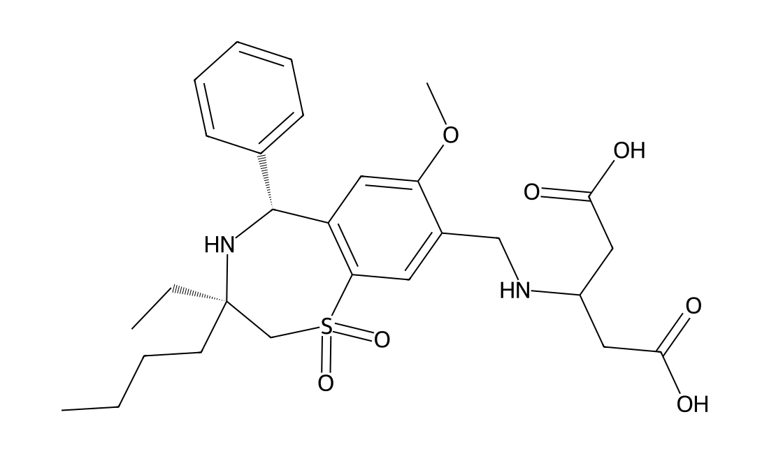
GSK2330672 is a highly potent, nonabsorbable ASBT inhibitor with excellent aqueous solubility, selectivity, and developability properties for evaluation in safety studies and ultimately humans. GSK2330672 will be a valuable clinical tool for exploring the therapeutic utility of a nonabsorbable ASBT inhibitor for treatment of patients with type 2 diabetes. in vivo: GSK2330672 results in potent inhibition of ASBT and very low oral absorption in the rat. GSK2330672 shows potent mouse and rat ASBT activity and was stable in GI stability assays. GSK2330672 is stable in the rodent GI tract and potently induced fecal bile acid excretion in mice, leading us to select these three compounds for mechanistic and efficacy studies in vivo in lean rats and Zucker Diabetic Fatty (ZDF) rats, respectively.
CAS 1079043-81-4
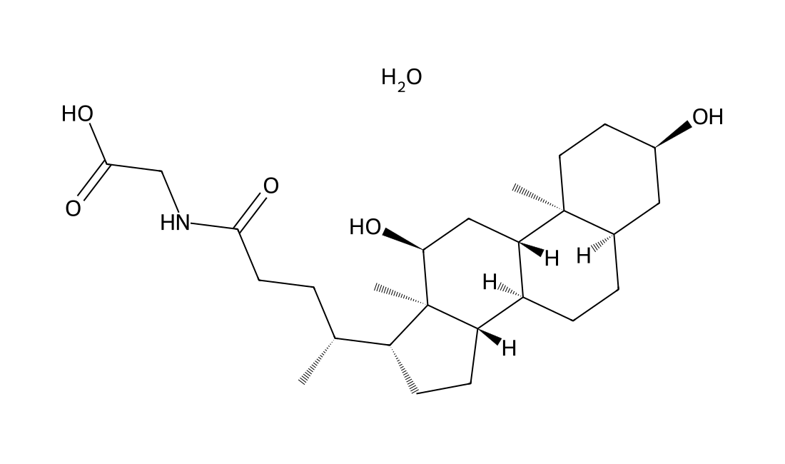
Glycodeoxycholic acid monohydrate is a nuclear receptor ligand.
CAS 93479-97-1
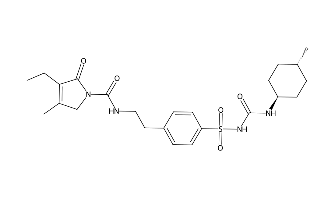
Glimepiride(Amaryl) is a medium-to-long acting sulfonylurea anti-diabetic compound with an ED50 of 182 μg/kg.Glimepiride (Hoe 490) is a new sulfonylurea. After oral administration of Hoe 490 to rabbits, blood glucose was lowered 3.5 times more than after glibenclamide (HB 419) and after intravenous administration, 2.5 times more. glimepiride decreased extracellular Aβ40 and Aβ42 levels. glimepiride may serve as a promising drug for the treatment of AD associated with diabetes. glimepiride was generally associated with lower risk of hypoglycemia and less weight gain compared to other sulfonylureas. Glimepiride use may be safer in patients with cardiovascular disease because of its lack of detrimental effects on ischemic preconditioning.
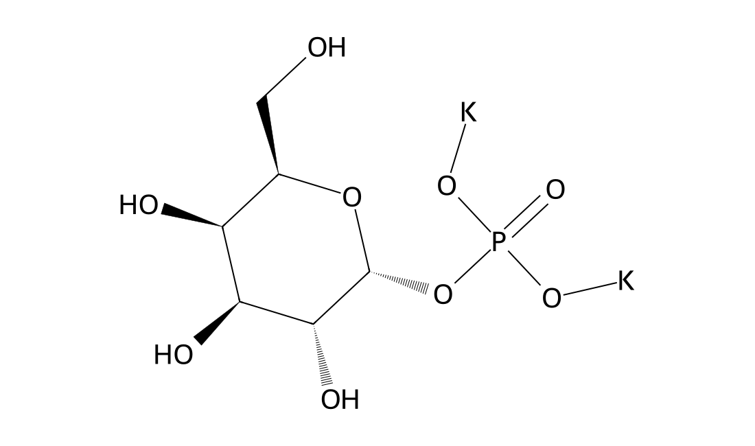
CAS 19046-60-7
Galactose 1-phosphate Potassium salt is is an intermediate in the galactose metabolism and nucleotide sugars
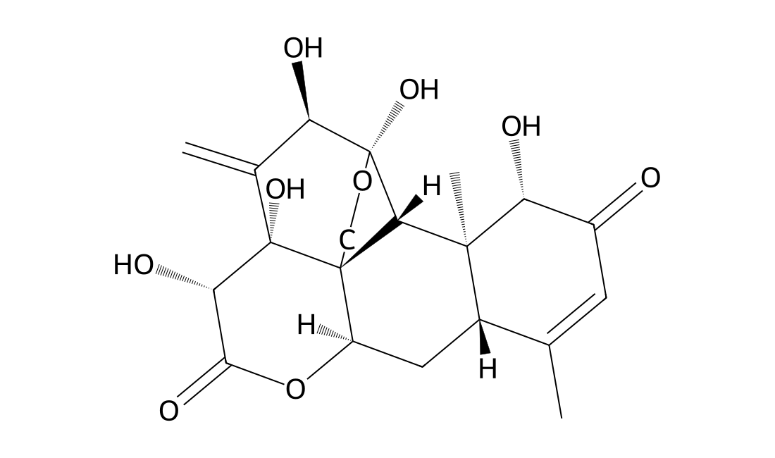
CAS 84633-29-4
In Vitro Eurycomanone (EN) significantly increased testosterone production dose-dependently at 0.1, 1.0 and 10.0 μM, but the two lower doses when combined with 3-isobutyl-1-methylxanthine (IBMX), the phosphodiesterase inhibitor were not significantly higher than EN or IBMX alone, except at a higher concentration. The molecular docking studies indicated EN and IBMX were binding at different sites of the enzyme. EN has no reversal of inhibition by aminoglutethimide, ketoconazole or nifedipine at the respective steroid oogenesis enzyme. The quassinoid was also non-responsive to the inhibition of oestrogen receptor by tamoxifen, but displayed improved for mestane inhibition of aromatase in reducing oestrogen production. The molecular docking studies further supported that EN and formestane bound to aromatase with similar orientations and free energy binding values.
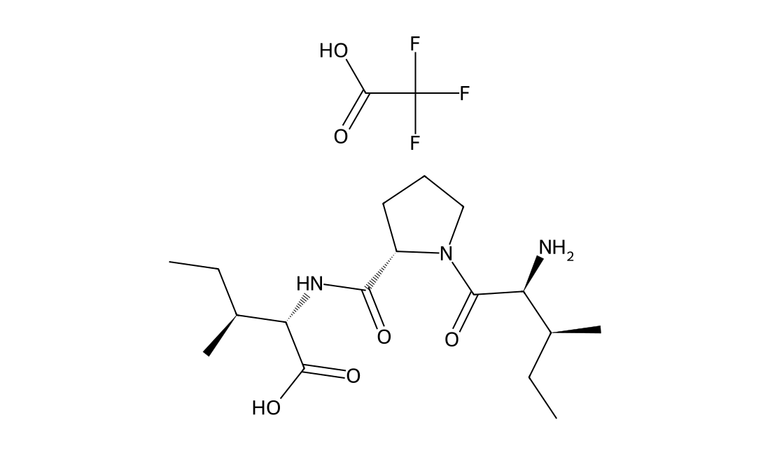
CAS 209248-71-5
In Vitro Diprotin A increases the phosphorylation of Src and vascular endothelial-cadherin (VE-cadherin) in human endothelial cells and disrupts endothelial cell-to-cell junctions, which are attenuated by CXCR4 (receptor of SDF-1α)-blocker or Src-inhibitor. In Vivo In the model of retinopathy of prematurity, Diprotin A increases not only retinal vascularity but also leakage. Additionally, in the murine diabetic retinopathy model, Diprotin A increases the phosphorylation of Src and VE-cadherin and aggravates vascular leakage in the retinas. Collectively, Diprotin A induces vascular leakage by augmenting the SDF-1α/CXCR4/Src/VE-cadherin signaling pathway.
CAS 81-13-0
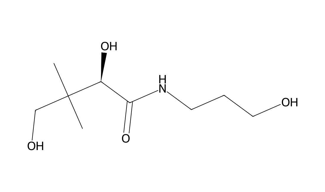
Dexpanthenol is an alcoholic analogue of D-pantothenic acid and cholinergic agent.
CAS 173865-33-3
CDD3505 specifically induces hepatic cytochrome P450IIIA produces significant increases in HDL cholesterol.
CAS 939390-99-5
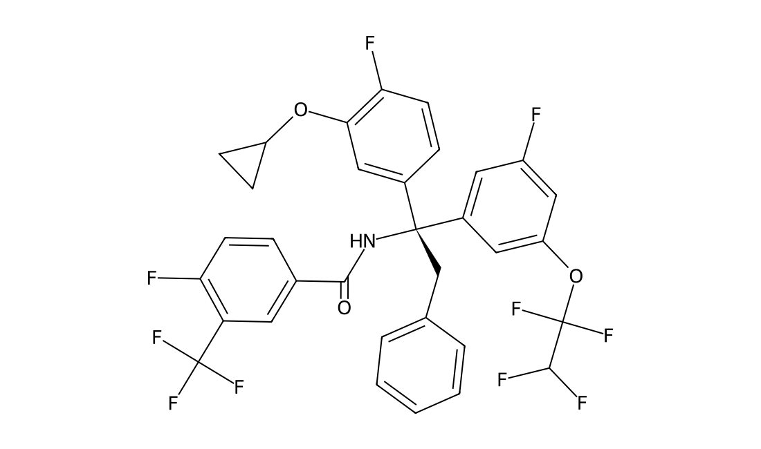
BMS-795311 is a potent and orally available CETP inhibitor with IC50 of 3.8 nM, inhibits cholesteryl ester (CE) transfer with IC50 of 0.22 uM; inhibits CE transfer activity at an oral dose of 1 mg/kg in human CETP/apoB-100 dual transgenic mice and increased HDL cholesterol content and size comparable to Torcetrapib in moderately-fat fed hamsters.
CAS 739366-20-2
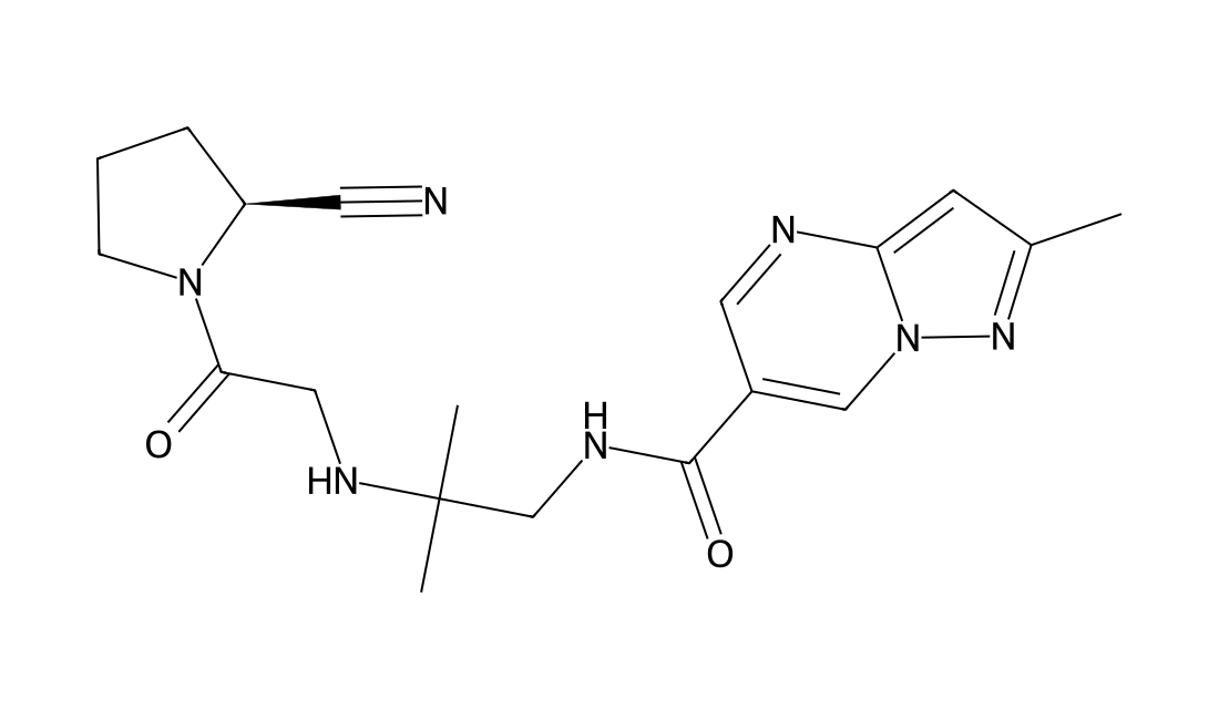
Anagliptin is a potent and selective DPP-4 inhibitor(IC50= 3.8 nM); > 10 fold less potent for DPP-8 and DPP-9. in vitro: In THP-1 cells, anagliptin reduced lipopolysaccharide-induced TNF-α production with inhibiting ERK phosphorylation and nuclear translocation of nuclear factor-κB . lipopolysaccharide (LPS)- or palmitate-induced elevations of inflammatory cytokine mRNA expressions in RAW264.7 macrophages were shown to be significantly suppressed by co-incubation with a DPP-4 inhibitor, anagliptin (10μM), despite low DPP-4 expression in the RAW264.7 cells. Anagliptin also acted on 3T3-L1 adipocytes, weakly suppressing the inflammatory cytokine expressions induced by LPS and TNF-α. When 3T3-L1 and RAW cells were co-cultured and stimulated with LPS, the effects of anagliptin on the suppression of cytokine expressions in 3T3-L1 adipocytes were more marked, and became evident at the 10μM concentration. in vivo: Treatment with anagliptin for 16 wk significantly reduced accumulation of monocytes and macrophages in the vascular wall, SMC content in plaque areas, and oil red O-stained area around the aortic valve without affecting glucose tolerance or body weight. Quantitative analysis also showed that anagliptin reduced the area of atherosclerotic lesion in apoE-deficient mice. Anti-inflammatory effects of anagliptin were also observed in vivo on the elevated hepatic and adipose expressions and serum concentrations of inflammatory cytokines in association with the suppression of hepatic NF-κB transcriptional activity, in LPS-infused mice.
CAS 18422-05-4
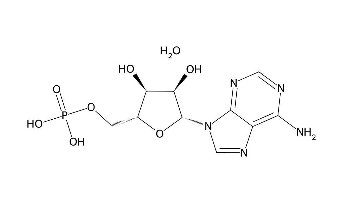
In Vitro Adenosine 5'-monophosphate monohydrate is an adenosine A1 receptor agonist[1]. Adenosine 5'-monophosphate monohydrate (5′-AMP)-induced cell toxicity is negligible at concentrations of 25 to 400 μM in RAW264.7 cells. Adenosine 5'-monophosphate monohydrate significantly attenuates the mRNA expression of tumor necrosis factor-α (TNF-α) and interleukin (IL)-6 in RAW264.7 cells. The dose-dependence of TNF-α and IL-6 mRNA show that at concentration of 400 μM, Adenosine 5'-monophosphate monohydrate exhibits the maximum inhibition. Exposure of cells to Adenosine 5'-monophosphate monohydrate significantly reduces recruitment of NF-κB p65 to TNF-α, IL-6, and IL-1β gene promoters. In Vivo C57BL/6J mice treated with Adenosine 5'-monophosphate monohydrate (5′-AMP) markedly increases hepatic adenosine level. Survival rate in PBS-treated mice (n=15) is 60% (8 h) and 33.3% (24 h), whereas survival rate in Adenosine 5'-monophosphate monohydrate-treated mice (n=15) is 100% (8 h) and 93.3% (24 h). Serum aspartate transaminase (AST) and alanine transaminase (ALT) levels are significantly lowered in the Adenosine 5'-monophosphate monohydrate group than in the vehicle group. The area and extent of necrosis is attenuated and the infiltration of inflammatory cells is reduced in the Adenosine 5'-monophosphate monohydrate group.
CAS 623-58-5
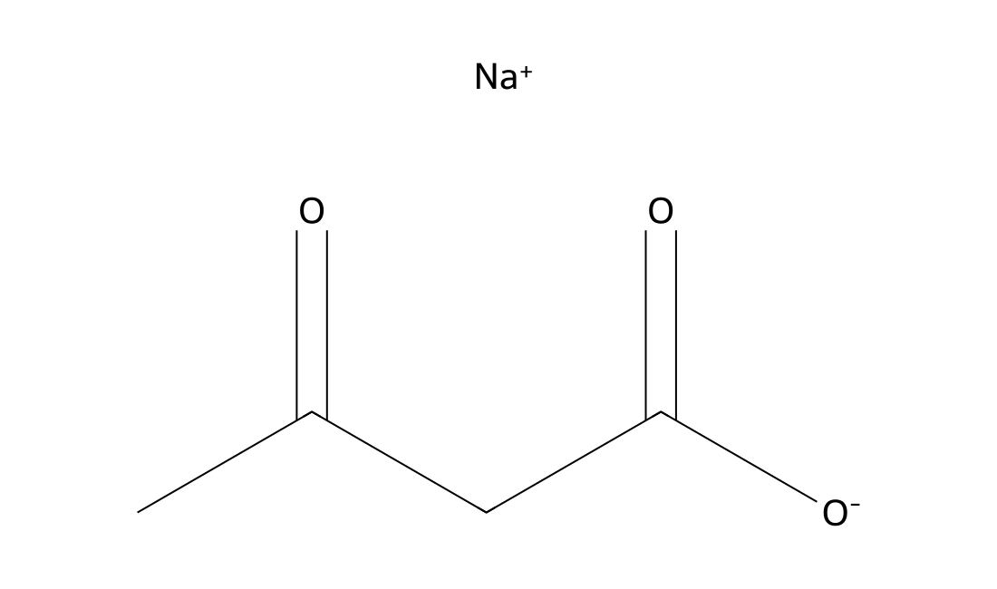
In Vitro Acetoacetic acid sodium salt induces oxidative stress to inhibit the assembly of very low density lipoprotein in bovine hepatocytes. Acetoacetic acid (0.6, 2.4, 4.8 mM) increases malondialdehyde (MDA) content in high-dose group (GH) group, decreases mRNA expression of Mn SOD, Cu/Zn SOD, and glutathione peroxidase (GSH-Px) in all groups, lowers catalase (CAT) mRNA expression in medial-dose group (GM), and GH groups. In addition, Acetoacetic acid down-regulates the mRNA expression of apolipoprotein B100 (ApoB100), apolipoprotein E (ApoE), and low density lipoprotein receptor (LDLR). Thus, VLDL assembly is decreased andtriglyceride (TG) accumulation is increased in these bovine hepatocytes.
CAS 63333-35-7
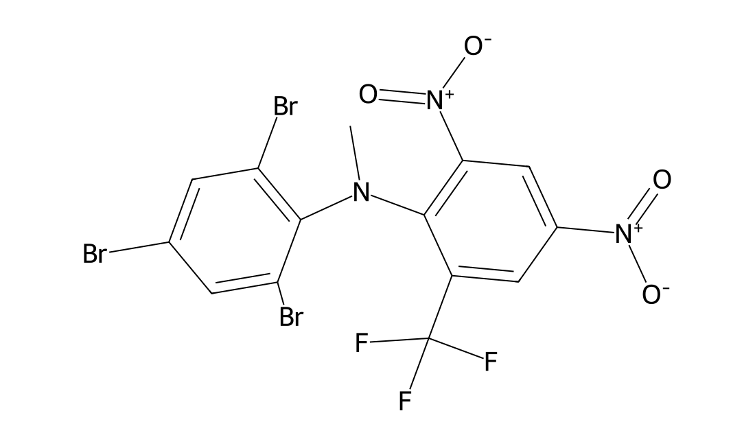
A rodenticide that poisons the central nervous system by uncoupling mitochondrial oxidative phosphorylation, which causes a decrease in adenosine triphosphate (ATP) synthesis.
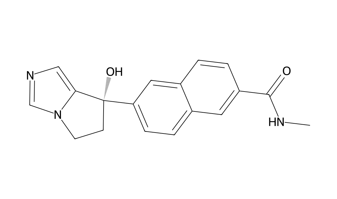
CAS 566939-85-3
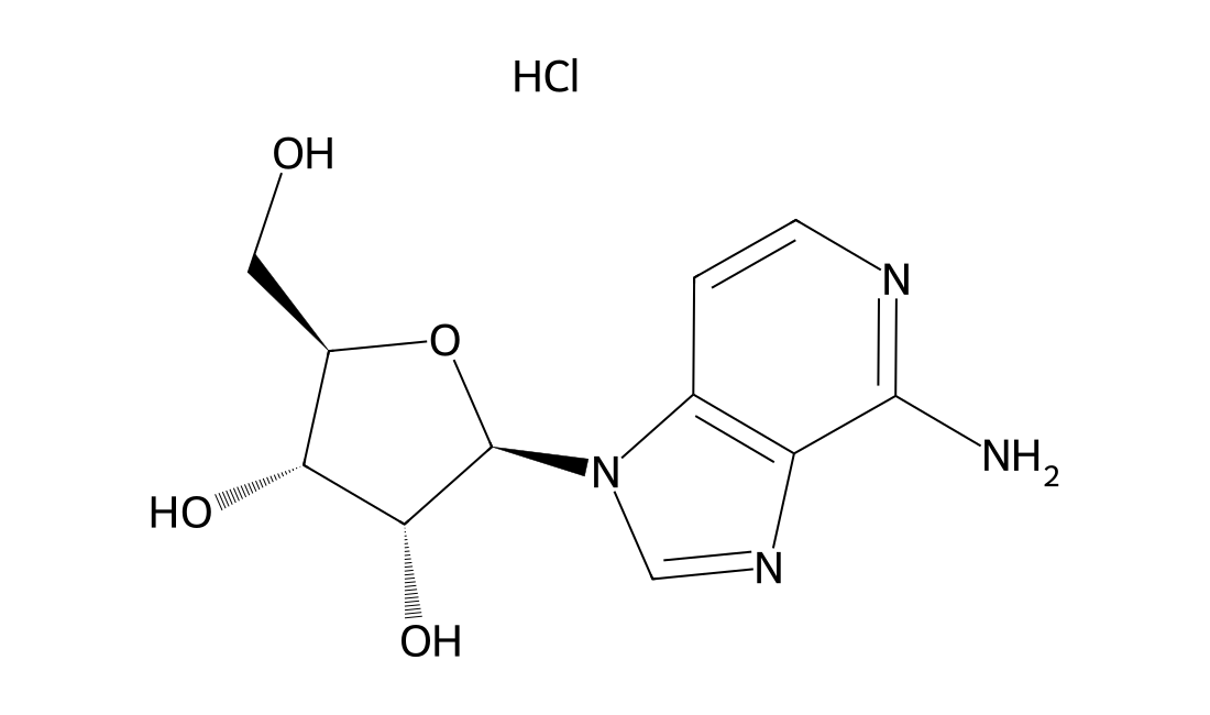
A highly selective CYP17A1 (17,20-lyase) inhibitor with IC50 of 17 nM for human 17,20-lyase; shows no activity on CYP3A4 (IC50>10 uM) and CYP11B1 (IC50>1 uM); reduces both serum testosterone and dehydroepiandrosterone concentrations in monkey model at a dose of 1mg/kg.Prostate cancerPhase 3 Discontinued
CAS 86583-19-9
In Vitro 3-Deazaadenosine is an inhibitor of S-adenosylhomocysteine hydrolase, with a Ki of 3.9 µM. 3-Deazaadenosine shows anti-HIV effect, and inhibits p24 antigen in peripheral blood mononuclear (PBMCs) cells infected with HIV-1 isolates (A012 and A018) with IC50s of 0.15 and 0.20 µM, respectively. 3-Deazaadenosine (1-100 µM) inhibits LPS-induced expression of TNF-α mRNA, increases DNA binding activity of NF-κB, and causes proteolytic degradation of IκBα, but Not IκBβ in RAW 264.7 cells. 3-Deazaadenosine (100 µM) enhances nuclear translocation of NF-κB, but blocks LPS-induced NF-κB transcriptional activity, and such inhibition is augmented by the addition of homocysteine. 3-Deazaadenosine (50, 100 µM) dose-dependently inhibits the phosphorylation of Raf and ERK, protein-dependent kinase 1, protein kinase B (Akt), and forkhead transcription factor FoxO1a. 3-Deazaadenosine (50 µM) suppresses vascular smooth muscle cell (VSMC) proliferation via interfering with Ras signaling.