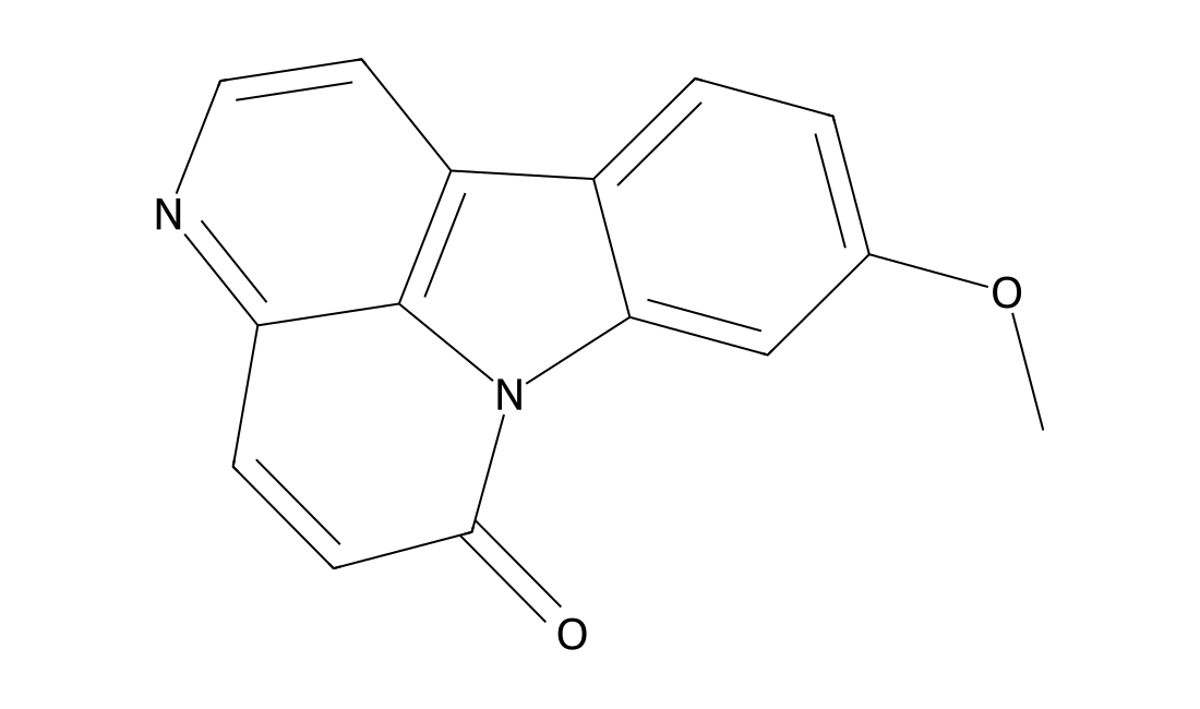
9-Methoxycanthin-6-one
CAS 74991-91-6
9-Methoxycanthin-6-one, a canthin-6-one alkaloid, is present in intact plant parts and in callus tissues of different explants. 9-Methoxycanthin-6-one shows anti-tumor activity.

Naturally occurring compounds for biological and chemical research.
Products may appear in more than one category. Counts refer to distinct catalog numbers within each category.
Other Research Tools (75) · Additional reagents, building blocks and specialized research tools.
Combine a keyword, research category and target to narrow the product list.
Loading catalog filters…
CAS 74991-91-6
9-Methoxycanthin-6-one, a canthin-6-one alkaloid, is present in intact plant parts and in callus tissues of different explants. 9-Methoxycanthin-6-one shows anti-tumor activity.
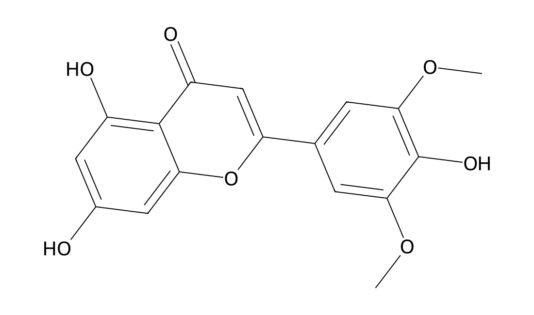
CAS 520-32-1
Tricin is a natural flavonoid present in large amounts in rice bran. Tricin can inhibit human cytomegalovirus (HCMV) replication by inhibiting CDK9. Tricin inhibits the proliferation and invasion of C6 glioma cells via the upregulation of focal-adhesion-finase (FAK)-targeting microRNA-7[1][2][3].
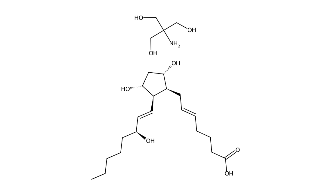
CAS 38562-01-5
Dinoprost(Prostaglandin F2α) tromethamine salt is a naturally occurring prostaglandin used in medicine to induce labor and as an abortifacient.
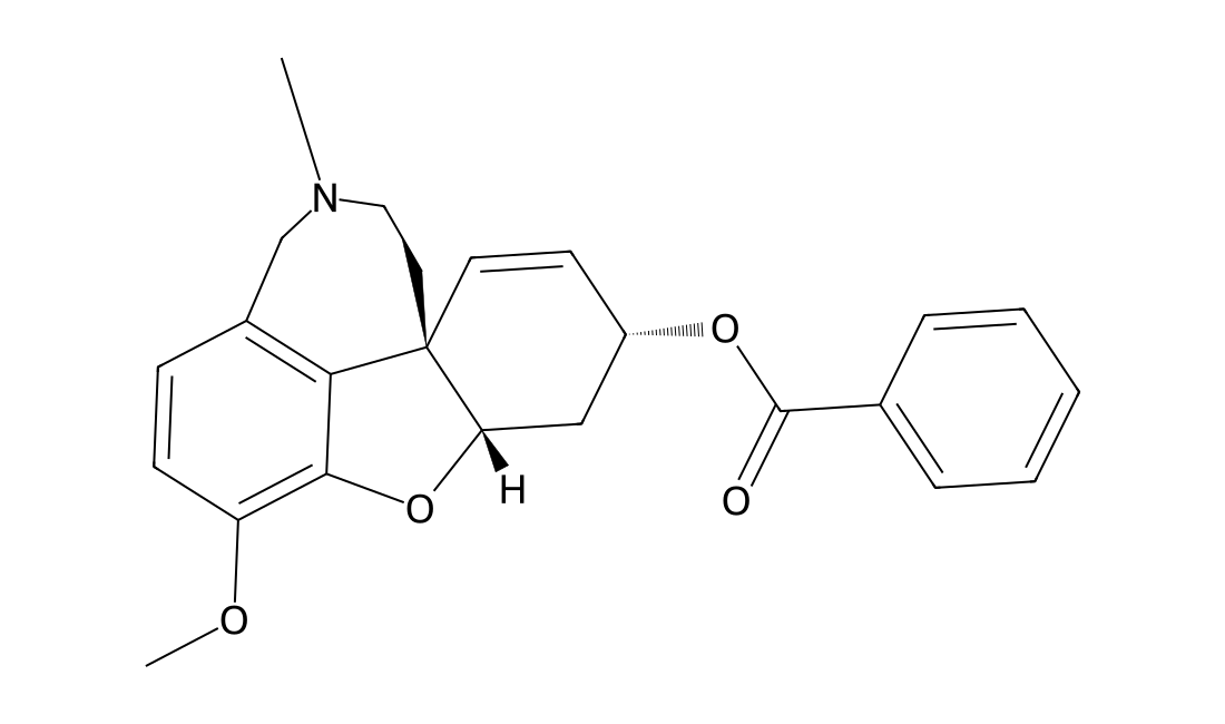
CAS 224169-27-1
GLN-1062 (Memogain) is an inactive pro-drug of galantamine, the latter being a plant alkaloid approved for the treatment of mild to moderate Alzheimer's disease.
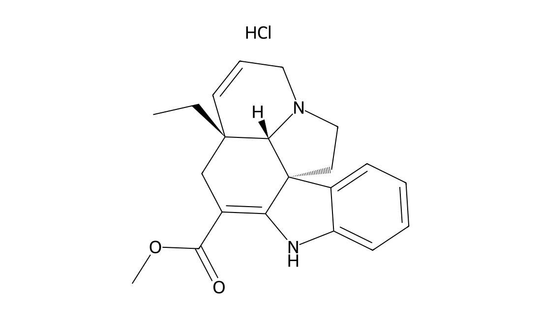
CAS 29479-00-3
Tabersonine is an indole alkaloid mainly isolated from Catharanthus roseus. Tabersonine disrupts Aβ(1-42) aggregation and ameliorates Aβ aggregate-induced cytotoxicity. Tabersonine has anti-inflammatory activities and is a potential therapeutic candidate for the treatment of ALI/ARDS[1].
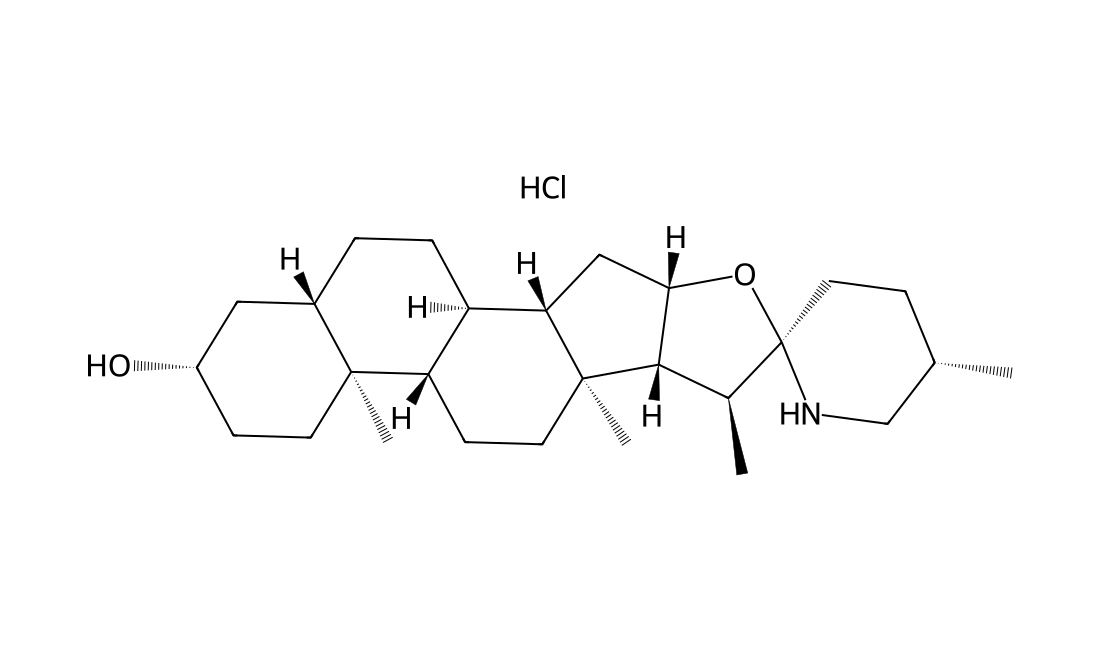
CAS 6192-62-7
Tomatidine hydrochloride acts as an anti-inflammatory agent by blocking NF-κB and JNK signaling.
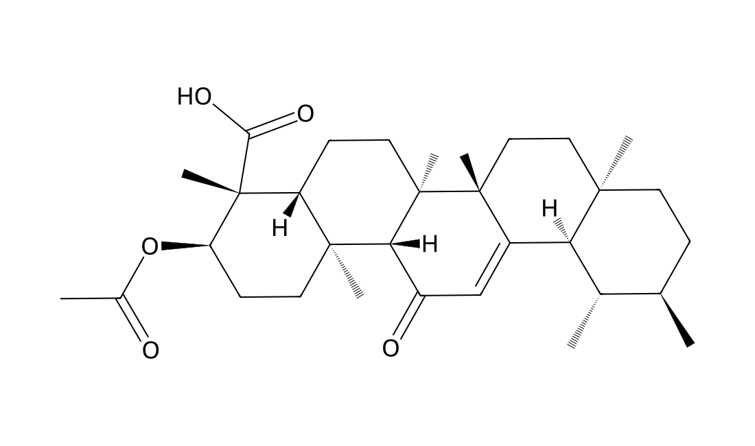
CAS 67416-61-9
Acetyl-11-Keto-β-Boswellic Acid (AKBA) is an active triterpenoid compound from the extract of Boswellia serrate; a novel Nrf2 activator.
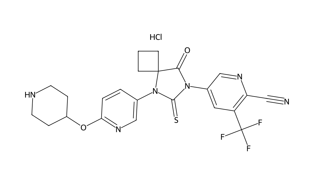
CAS 2110428-64-1
JNJ63576253, also known as TRC253, is a potent and orally active androgen receptor antagonist. TRC253 specifically binds to both wild-type and certain mutant forms of AR, thereby preventing androgen-induced receptor activation and facilitating the formation of inactive complexes that cannot translocate to the nucleus.
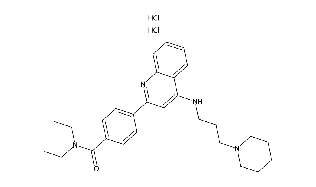
CAS 2310135-46-5
YUN35454, also known as LMPTP INHIBITOR 1 is a selective inhibitor of low molecular weight protein tyrosine phosphatase (LMPTP), with an IC50 of 0.8 μM LMPTP-A. LMPTP is a key promoter of insulin resistance and that LMPTP inhibitors would be beneficial for treating type 2 diabetes. YUN35454 was first reported in Nature Chemical Biology (2017), 13(6), 624-632.
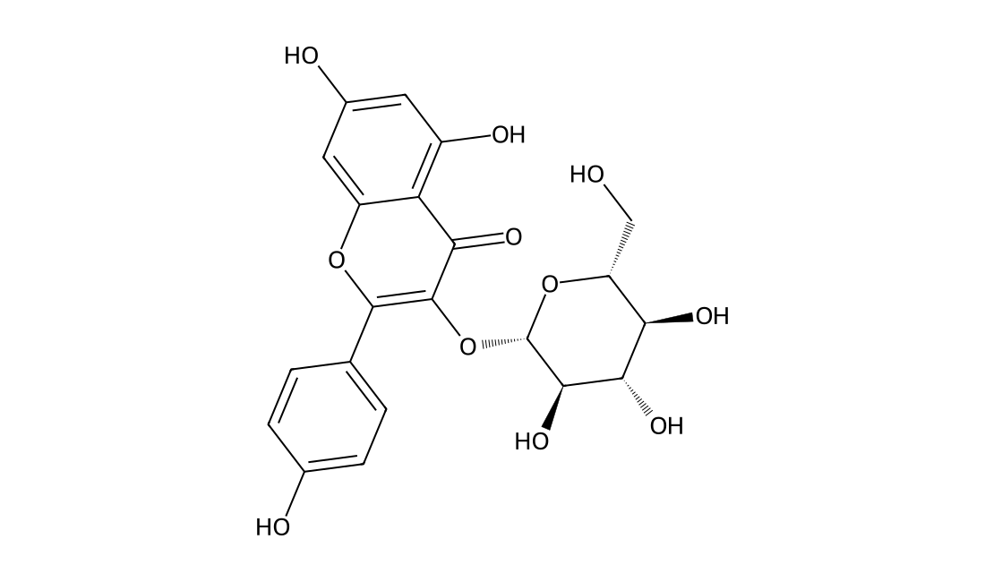
CAS 480-10-4
Astragalin (kaempferol-3-O-glucoside) is a flavonoid with anti-inflammatory activity and newly found in persimmon leaves and green tea seeds.IC50 value:Target: in vitro: Astragalin nontoxic at ≤ 20 μM suppressed cellular induction of Toll-like receptor 4 (TLR4) and ROS production enhanced by LPS. Both LPS and H2O2 induced epithelial eotaxin-1 expression, which was blocked by astragalin. LPS activated and induced PLCγ1, PKCβ2, and NADPH oxidase subunits of p22phox and p47phox in epithelial cells and such activation and induction were demoted by astragalin or TLR4 inhibition antagonizing eotaxin-1 induction. H2O2-upregulated phosphorylation of JNK and p38 MAPK was dampened by adding astragalin to epithelial cells, while this compound enhanced epithelial activation of Akt and ERK. H2O2 and LPS promoted epithelial apoptosis concomitant with nuclear condensation or caspase-3 activation, which was blunted by astragalin [1]. astragalin suppressed the expression of tumor necrosis factor α, interleukin 6, and nitric oxide in a dose-dependent manner in mMECs [2]. astragalin attenuated the infiltration of inflammatory cells, the activity of myeloperoxidase (MPO) and the expression of tumor necrosis factor-α (TNF-α), interleukin-6 (IL-6) and interleukin-1β (IL-1β) in a dose-dependent manner. Additionally, Western blotting results showed that astragalin efficiently blunt decreased nuclear factor-kappaB (NF-κB) activation by inhibiting the degradation and phosphorylation of IκBα and the nuclear translocation of p65 [3]. Astragalin significantly reduced LPS-induced expression of iNOS, COX-2 and cytokines/chemokines, and production of NO in J774A.1 mouse macrophages. Astragalin inhibited LPSinduced activation of NF-κB as indicated by inhibition of degradation of IκBα, nuclear translocation of NF-κB, and NF-κB dependent gene reporter assay [4].in vivo: Mice were injected intraperitoneally (i.p.) with lipopolysaccharide (LPS) (dose range: 5-40 mg/kg). pretreatment with astragalin can improve survival during lethal endotoxemia and attenuate inflammatory responses in a murine model of lipopolysaccharide-induced acute lung injury [4].
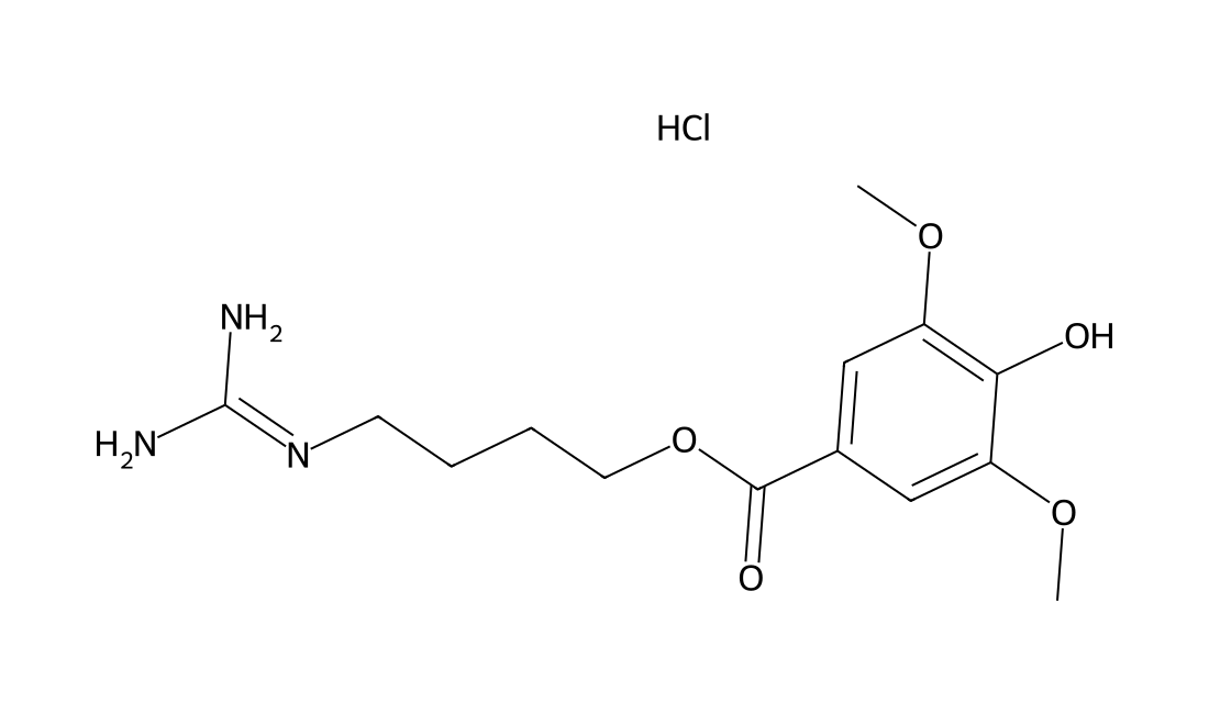
CAS 24735-18-0
Leonurine hydrochloride is an alkaloid isolated from Herba leonuri, with anti-oxidative and anti-inflammatory.
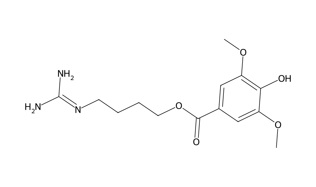
CAS 24697-74-3
Leonurine is an alkaloid isolated from Herba leonuri, with anti-oxidative and anti-inflammatory.
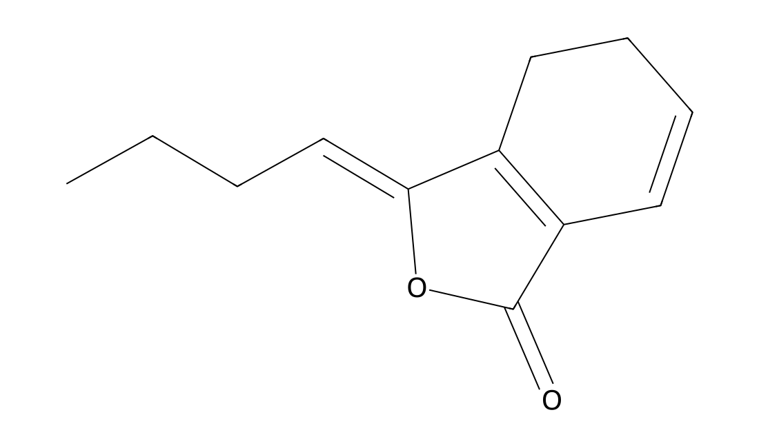
CAS 4431-01-0
Ligustilide is an effective constituent extracted from Angelica sinensis.IC50 value:Target:In vitro: To investigate the neuroprotective of ligustilide (LIG) against glutamate-induced apoptosis of PC12 cells, cell viability were examined by MTT assay. Pretreatment with ligustilide (1, 5, 15 μmol · L(-1)) significantly improved cell viability. The apoptosis rate in glutamate-induced PC12 cells was 13.39%, and decreased in the presence of ligustilide (1, 5, 15 μmol · L(-1)) by 9.06%, 6.48%, 3.82%, separately. Extracellular accumulation of Ca2+ induced by glutamate were significantly reduced by ligustilide [1].
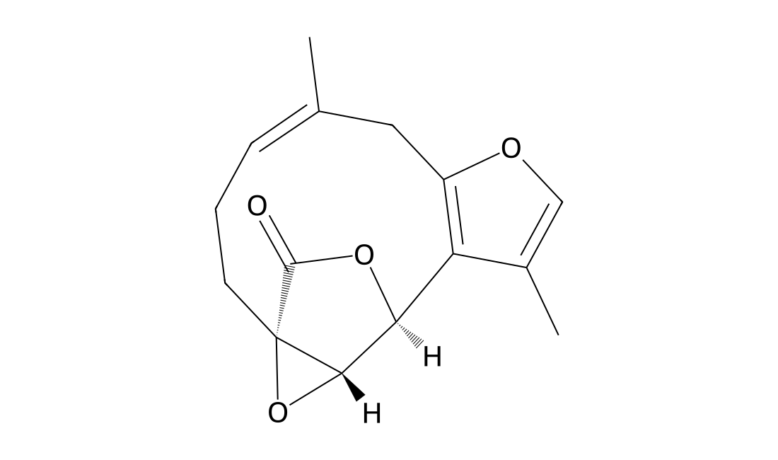
CAS 13476-25-0
Linderane, isolated from the root of Lindera strychnifolia, is an irreversible inhibitor cytochrome P450 2C9 (CYP2C9). Linderane has the potential to relieve pain and cramp[1].
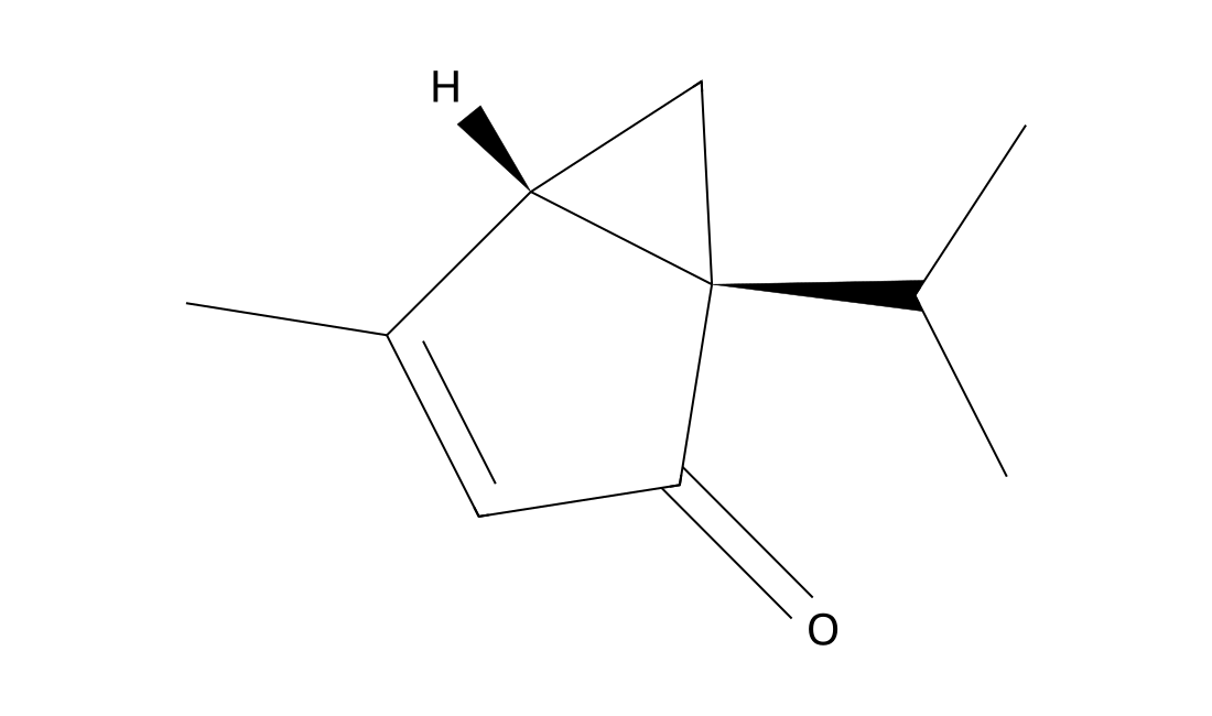
CAS 546-78-1
Umbellulone is an active constituent of the leaves of Umbellularia californica. Umbellulone stimulates the TRPA1 channel in a subset of peptidergic, nociceptive neurons, activating the trigeminovascular system via this mechanism[1].
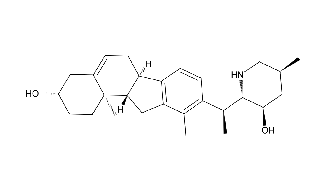
CAS 60-70-8
Veratramine(NSC17821; NSC23880) is useful as a signal transduction inhibitor for treating tumors.
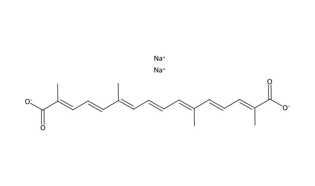
CAS 591230-99-8
In Vitro Transcrocetinate (Transcrocetin, trans-Crocetin), a saffron metabolite originating from the crocin apocarotenoids, has been shown to exert strong NMDA receptor affinity and is thought to be responsible for the CNS activity of saffron.To ensure unchanged viability of Caco-2 cells throughout the transport experiments, cellular mitochondrial dehydrogenase activity of Caco-2 cells is measured by MTT assay after a 24 h incubation period with the test compounds: Hydroalcoholic saffron extract saffron extract (SE, 0.5-1 mg/mL) and crocin-1 (250-1000 µM) reveal no negative significant changes in cellular viability. Transcrocetinate at 10 µM level does not change viability while higher concentrations (40-160 µM) reduces significantly cellular viability.
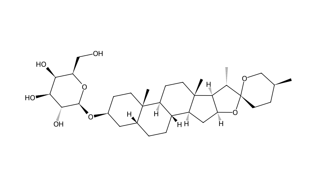
CAS 68422-00-4
Timosaponin A1 is a coprostane type steroidal saponin isolated from Rhizoma Anemarrhenae.
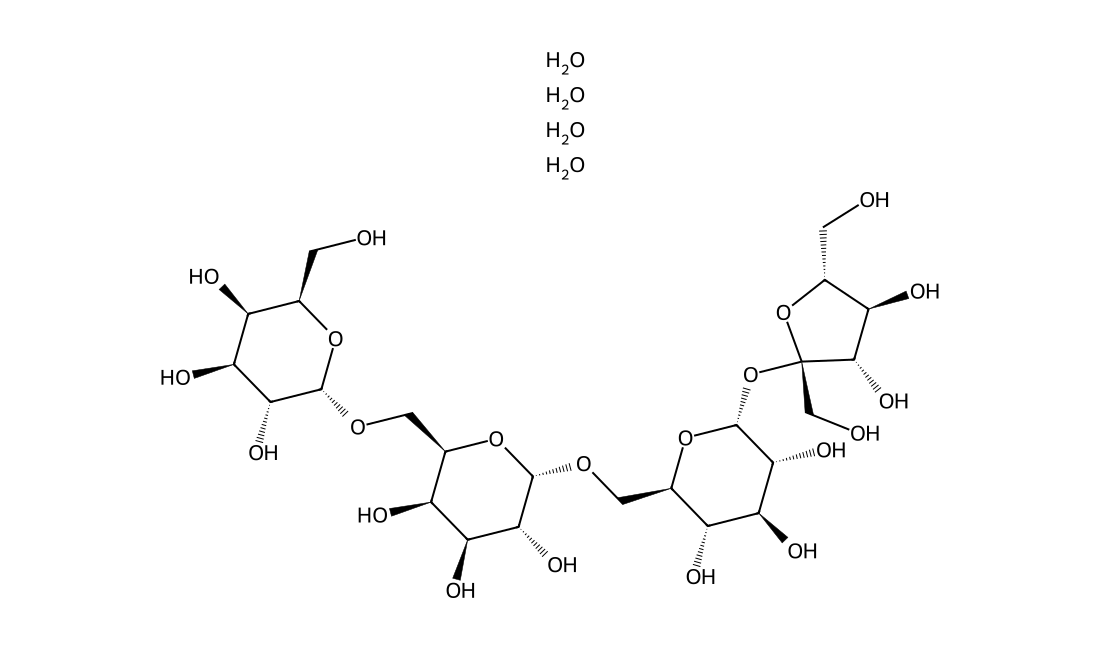
CAS 10094-58-3
In Vitro Stachyose highly promotes proliferation of lactic acid bacteria (LAB) by inducing LAB to produce more α-galactosidase to hydrolyze stachyose. In Vivo Stachyose may improve bioavailability of genistein via inhibiting intestinal degradation and first-pass metabolism of soy genistein.
CAS 96574-01-5
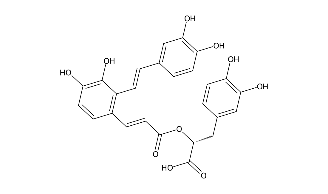
Salvianolic acid A is cell protective antioxidant originally isolated from Salvia. It has been found to attenuate PDGF-induced ROS formation in hepatic stellate cells and protected against isoproterenol-induced myocardial damage in rats. Salvianolic acid A acts as scavengers of reactive oxygen species, reduces the adherence of leukocytes to endothelial cells, inhibits matrix metalloproteinases and inflammation. It has cardioprotective effects as it suppresses apoptosis, reduces lipid peroxidation in damaged cardiac tissue and decreases the leakage of lactic acid dehydrogenase. For the detailed information of Salvianolic acid A, the solubility of Salvianolic acid A in water, the solubility of Salvianolic acid A in DMSO, the solubility of Salvianolic acid A in PBS buffer, the animal experiment(test) (test) of Salvianolic acid A, the cell expriment (test) of Salvianolic acid A, the in vivo, in vitro and clinical trial test of Salvianolic acid A, the EC50, IC50,and Affinity of Salvianolic acid A,, please contact DC Chemicals.
CAS 1311473-28-5
Perisesaccharide C is an oligosaccharide isolated from the root barks of Periploca sepium.
CAS 1095261-93-0
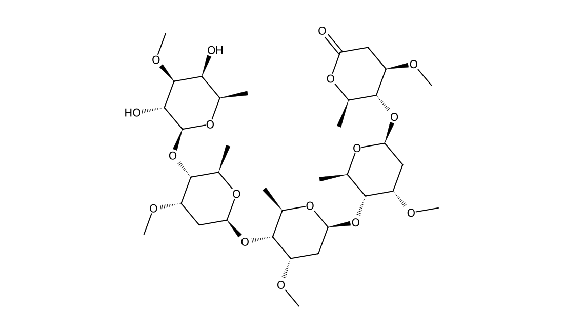
Perisesaccharide B is an oligosaccharide isolated from the root barks of Periploca sepium.
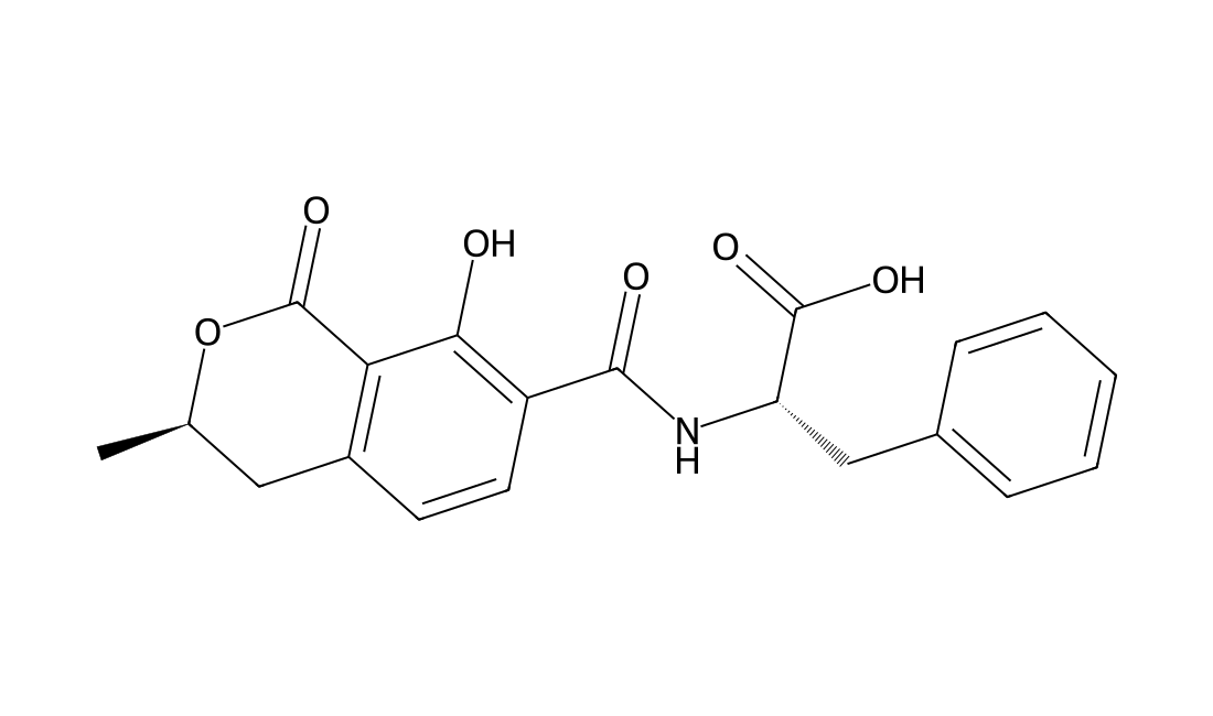
CAS 4825-86-9
Ochratoxin B(OTB) is a non-chlorinated analog of OTA that has cytotoxic effects on kidney and liver cells in vitro but only minor effects in vivo, due to its rapid metabolism and excretion.OTB inhibits cell proliferation of human liver HepG2 cells at doses as low as 1 µg/ml but lacks the genotoxic activity of OTA, even at higher concentrations.
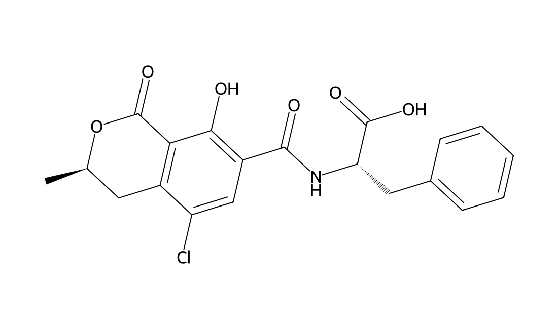
CAS 303-47-9
Ochratoxin A is a mycotoxin isolated from the Aspergillus ochraceus mold, a common fungal presence on moldy food. Ochratoxin A demonstrates nephrotoxicity and teratogenesis in animals, and shows inhibition of bacterial, yeast, and liver FARSL (phenylalanyl-tRNA synthetases) competitive with phenylalanine. As these activities are partially attenuated by phenylalanine, Ochratoxin A is suggested to produce its effects through interacting with proteins that recognize phenylalanine as a substrate, and has been demonstrated to inhibit PAH (phenylalanine hydroxylase). Derivatives of Ochratoxin A with other amino acid moieties substituted for the phenylalanine-mimic portion of the molecule demonstrate inhibition of the aminoacyl tRNA synthetases respective to those amino acids. Ochratoxin A is described to enhance lipid peroxidation and also present DNA single strand breaks, both suggesting oxidative damage as a partial mediator of Ochratoxin A cellular effects, and EPR spectroscopy with a spin trap agent identified the production of hydroxyl radicals by Ochratoxin A in the presence of NADPH. The nephrotoxic effects of Ochratoxin A are shown to be prevented by SOD (superoxide dismutase) and catalase, further indicating oxidative damage as the mediator of Ochratoxin A toxicity. Ochratoxin A is also demonstrated to increase ATP-dependent Ca2+ pump activity in renal cortex endoplasmic reticulum.
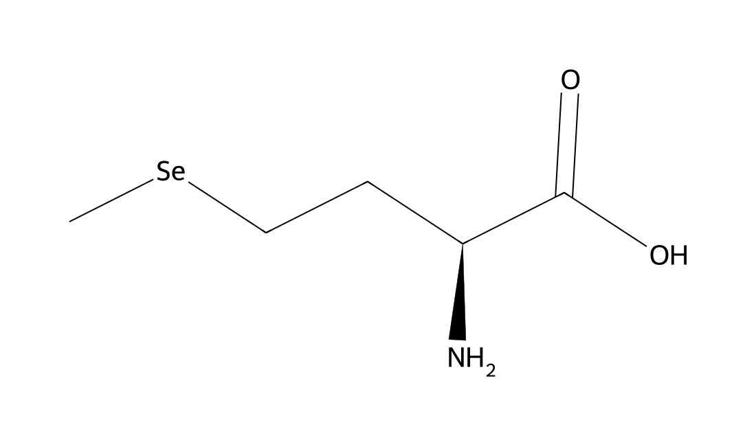
CAS 3211-76-5
L-SelenoMethionine is a major natural food-form of selenium.The median lethal dose (LD50) of L-SelenoMethionine in rats given an intraperitoneal injection was determined to 4.25 mg Se/kg body and thus is comparable to that of selenite or selenate. In mice, the LD50 of L-SelenoMethionine was 8.8 ± 1.37 mg Se/kg, and the minimal lethal dose, 4.0 mg Se/kg, after intravenous injection.
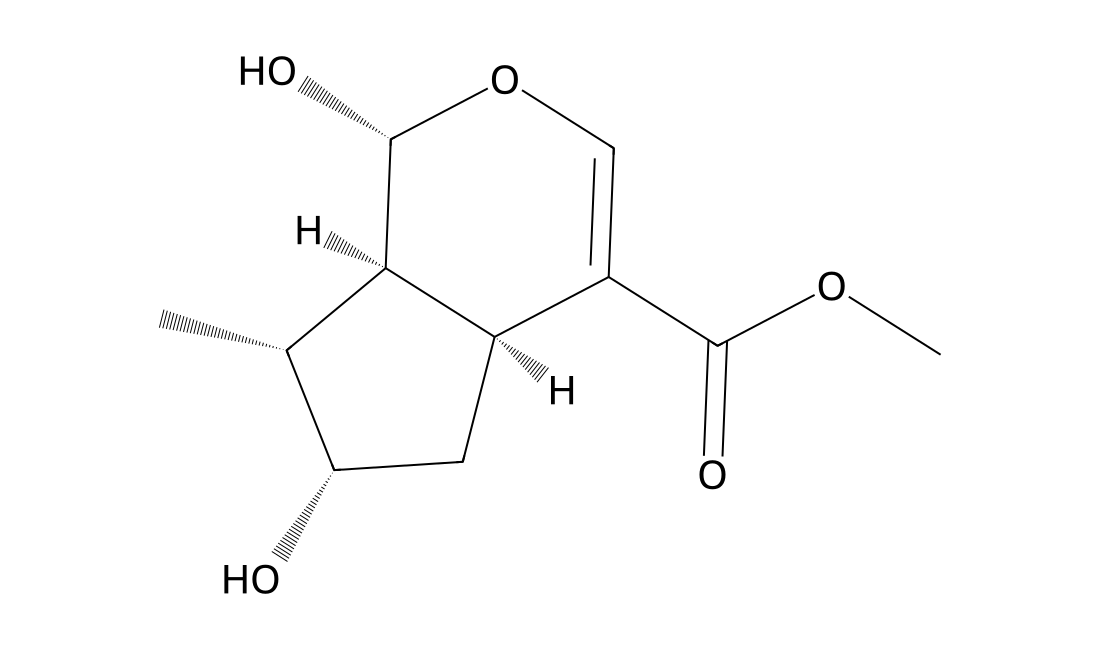
CAS 29748-10-5
In Vitro Although Loganetin does not possess antibacterial activity of its own, but in combination, it appreciably reduces the minimum inhibitory concentration (MIC) of nalidixic acid (NA) against nalidixic acid resistant (NAREC) and nalidixic acid sensitive (NASEC) strains of Escherichia coli. Loganetin, a very common, inexpensive, and non-toxic natural product may finds its application in the antibacterial drug development for treating multidrug-resistant Gram negative infections.
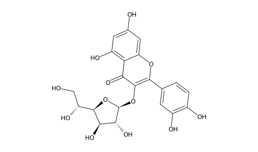
CAS 21637-25-2
In Vitro Isoquercitrin occurs widely in plants.10 μM of Isoquercitrin of the seven compounds partially but significantly blunt the negative effect of H2O2 on RGC-5 cells, as the viability of cells in the presence of H2O2 is increased from approximately 63% to 83% and 90% at 10 and 50 μM, respectively. Indeed, 50 μM Isoquercitrin is more effective as a neuroprotectant than the same concentration of EGCG. In Vivo In animals receiving Isoquercitrin or quercetin, eosinophil counts are lower in the BALF, blood and lung parenchyma. Neutrophil counts in blood and IL-5 levels in lung homogenate are lower only in Isoquercitrin-treated mice. No alterations in mononuclear cell numbers were observed.
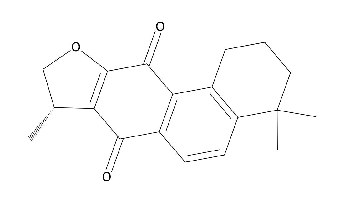
CAS 22550-15-8
In Vitro It is showed that tanshinones significantly inhibited A549 proliferation and Isocryptotanshinone (ICTS) exhibits the strongest activity. Isocryptotanshinone inhibits the constitutive STAT3 and p-STAT3 (Y705) expression in concentration and time-dependent manners. Isocryptotanshinone dramatically inhibits the IL-6-stimulated expression of p-STAT3 (Y705) in a time-dependent manner.
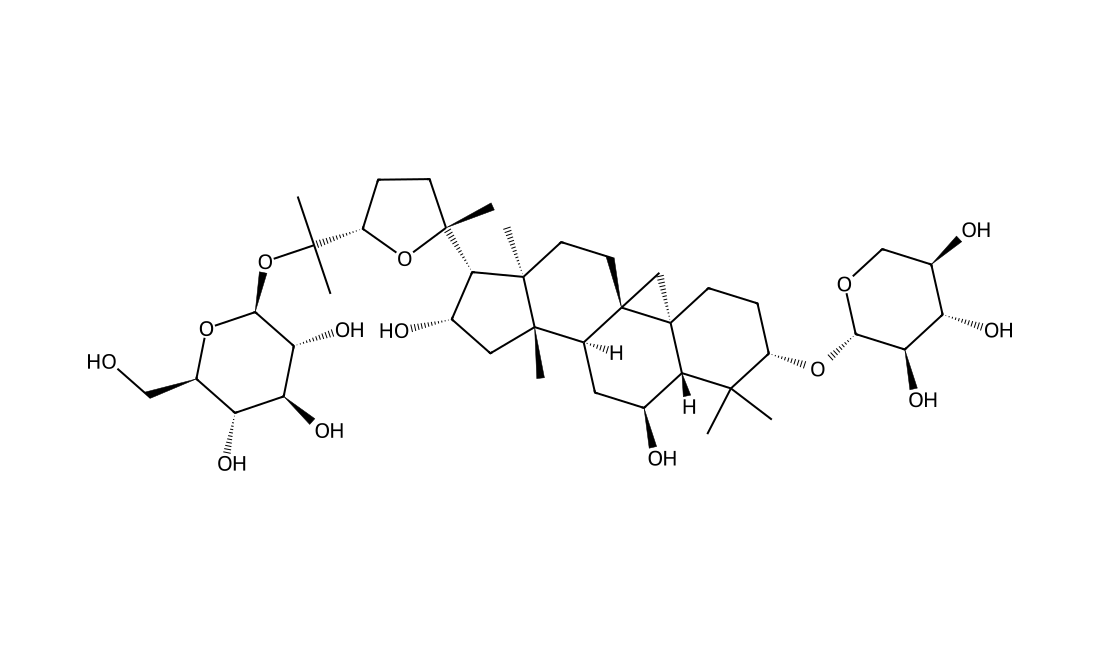
CAS 136033-55-1
Isoastragaloside IV is a triterpene oligoglycoside isolated from Astragali Radix.
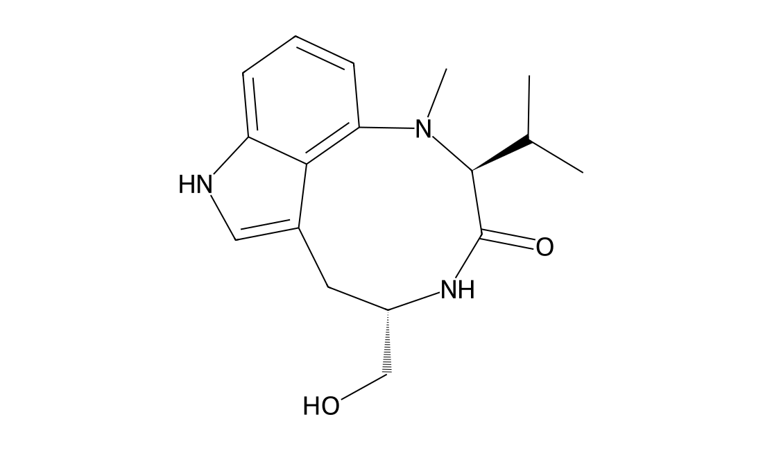
CAS 90365-57-4
Indolactam V ((-)-Indolactam V) is an indole alkaloid tumor promoter that activates protein kinase C (PKC), binds to PKC regulatory domains of mouse skin PKCη and rat brain PKCγ with Ki values of 3.4 nM and 1 uM, respectively; induces differentiation of human embryonic stem cells into pancreatic progenitors through activation of PKC signaling.
CAS 22419-74-5
Incensole is a diterpene originally isolated from frankincense produced by B. carterii.It inhibits degradation of inhibitor of NF-κB (IκBα) in a concentration-dependent manner in TNF-α-stimulated HeLa cells when used at concentrations ranging from 60 to 140 μM.
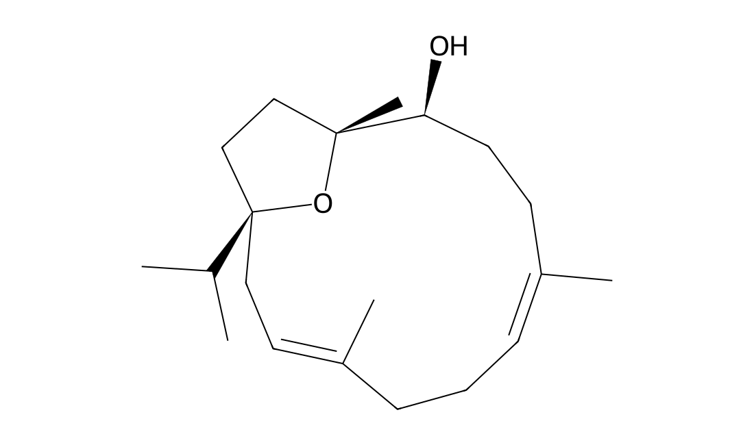
CAS 17680-84-1
Homoplantaginin shows IC50 of reduction level of DPPH radical at 0.35 μg/mL. In human hepatocyte HL-7702 cells exposed to H2O2, the addition of 0.1-100μg/mL of homoplantaginin significantly reduces lactate dehydrogenase leakage, and increases glutathione, glutathione peroxidase and superoxide dismutase in supernatant. Homoplantaginin (0.1, 1, 10 μM) dose-dependently reduces expression of toll-like receptor-4 evoked by palmitic acid (100 μM). Homoplantaginin tightly controlls palmitic acid-induced reactive oxygen species to prevent nucleotide-binding domain-like receptor 3 (NLRP3) inflammasome activation by suppressing reactive oxygen species-sensitive thioredoxin-interacting protein, NLRP3, and caspase-1. Pre-treatment of homoplantaginin on human umbilical vein endothelial cells significantly inhibits palmitic acid induced TNF-α and IL-6 mRNA expression, and IKKβ and NF-κB p65 phosphorylation. Homoplantaginin significantly modulates the Ser/Thr phosphorylation of IRS-1, improves phosphorylation of Akt and endothelial nitric oxide synthase, and increases NO production in the presence of insulin.Homoplantaginin shows IC50 of reduction level of DPPH radical at 0.35 μg/mL. In human hepatocyte HL-7702 cells exposed to H2O2, the addition of 0.1-100μg/mL of homoplantaginin significantly reduces lactate dehydrogenase leakage, and increases glutathione, glutathione peroxidase and superoxide dismutase in supernatant. Homoplantaginin (0.1, 1, 10 μM) dose-dependently reduces expression of toll-like receptor-4 evoked by palmitic acid (100 μM). Homoplantaginin tightly controlls palmitic acid-induced reactive oxygen species to prevent nucleotide-binding domain-like receptor 3 (NLRP3) inflammasome activation by suppressing reactive oxygen species-sensitive thioredoxin-interacting protein, NLRP3, and caspase-1. Pre-treatment of homoplantaginin on human umbilical vein endothelial cells significantly inhibits palmitic acid induced TNF-α and IL-6 mRNA expression, and IKKβ and NF-κB p65 phosphorylation. Homoplantaginin significantly modulates the Ser/Thr phosphorylation of IRS-1, improves phosphorylation of Akt and endothelial nitric oxide synthase, and increases NO production in the presence of insulin.
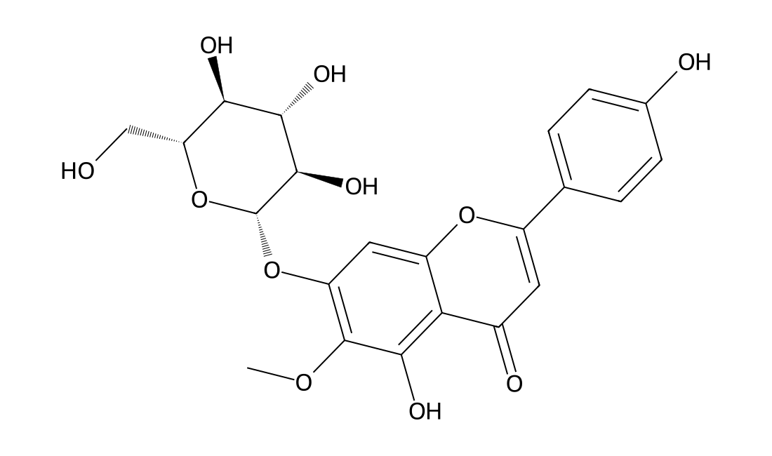
CAS 81525-13-5
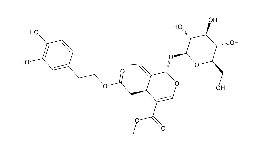
Forsythoside B concentration-dependently down-regulates the levels of TNF-α, IL-6 and high-mobility group-box 1 protein (HMGB1) in lipopolysaccharide (LPS)-stimulated RAW264.7 cells, inhibits the IκB kinase (IKK) pathway and modulated nuclear factor (NF)- κB.Intravenous injection of forsythoside B alone or plus imipenem reduces serum levels of TNF-α, IL-6, HMGB1, triggering receptor expressed on myeloid cells (TREM-1) and endotoxin, while the serum level of IL-10 is up-regulated and myeloperoxidase (MPO) in lung, liver and small intestine is reduced. Forsythoside B at doses higher than 8 mg/kg produces a significant neuroprotective potential in cerebral ischemia and reperfusion rats. Forsythoside B (20 mg/kg) demonstrates significant neuroprotective activity even after delayed administration at 1 h, 3 h and 5 h after cerebral ischemia and reperfusion. Forsythoside B 20 mg/kg attenuates histopathological damage as demonstrated by smaller brain infarct size and brain edema, decreased cerebral Evans blue extravasation and myeloperoxidase activity, inhibited cerebral phosphor-IκB-α and NF-κB expression. Forsythoside B shows a significant recovery in myocardial function with improvement of LVSP and +/-dp/dt(max). The myocardial infarct volume, serum levels of Tn-T, TNF-alpha and IL-6, content of MDA and MPO activity in myocardial tissue are all reduced, protein expression of HMGB1, phosphor-I kappaB-alpha and phosphor-NF-kappaB are down-regulated, while attenuate the decrease of SOD and GPx activities.
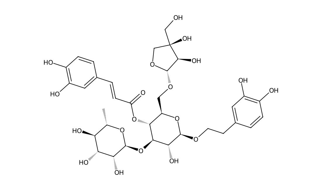
CAS 84633-29-4
In Vitro Eurycomanone (EN) significantly increased testosterone production dose-dependently at 0.1, 1.0 and 10.0 μM, but the two lower doses when combined with 3-isobutyl-1-methylxanthine (IBMX), the phosphodiesterase inhibitor were not significantly higher than EN or IBMX alone, except at a higher concentration. The molecular docking studies indicated EN and IBMX were binding at different sites of the enzyme. EN has no reversal of inhibition by aminoglutethimide, ketoconazole or nifedipine at the respective steroid oogenesis enzyme. The quassinoid was also non-responsive to the inhibition of oestrogen receptor by tamoxifen, but displayed improved for mestane inhibition of aromatase in reducing oestrogen production. The molecular docking studies further supported that EN and formestane bound to aromatase with similar orientations and free energy binding values.
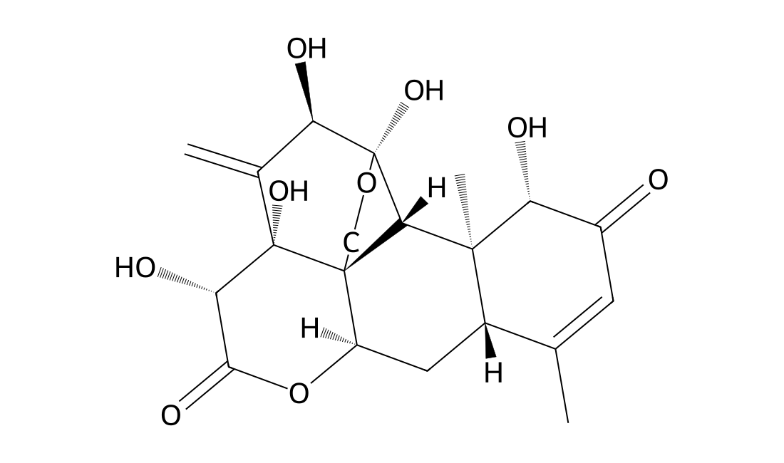
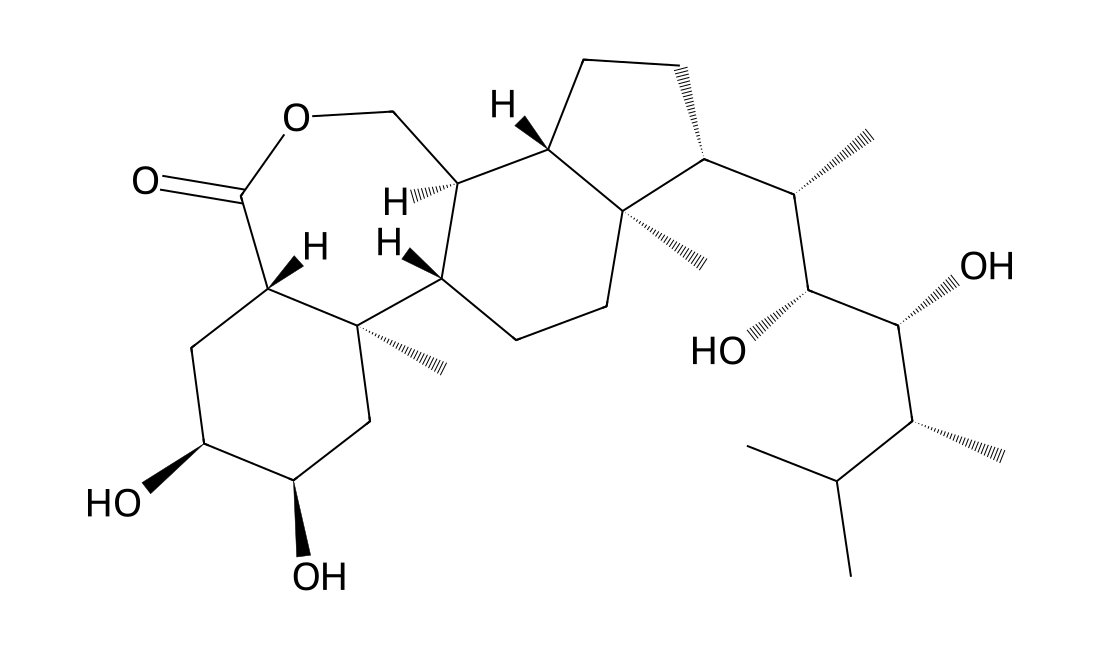
CAS 78821-43-9
Epibrassinolide (EBR; B1105; 24-Epibrassinolide) is a biologically active compound of the brassinosteroids, steroid-derived plant growth regulator family; was shown as a potential apoptotic inducer in various cancer cells without affecting the non-tumor cell growth. in vitro: Induction of apoptotic cell death was observed in prostate cancer cell lines after EBR treatment. In addition, EBR induced the decrease of intracellular polyamine levels, accompanied by a significant ornithine decarboxylase (ODC) down-regulation in each prostate cancer cell and also modulated ODC antizyme and antizyme inhibitor expression levels only in LNCaP cells. transcriptional expression of eight genes involved in astaxanthin biosynthesis exposed to EBR (25 and 50 mg/L) was analyzed using qRT-PCR. The results demonstrated that both 25 and 50 mg/L EBR could increase astaxanthin productivity and the eight carotenogenic genes were up-regulated by EBR with different expression profiles. Moreover, EBR25 induction had a greater influence on the transcriptional expression of ipi-1, ipi-2, crtR-B, lyc and crtO (> 5- fold up-regulation) than on psy, pds, bkt; EBR50 treatment had a greater effect on the transcriptional expression of ipi-2, pds, lyc, crtR-B, bkt and crtO than on ipi-1 and psy. Furthermore, astaxanthin biosynthesis under EBR was up-regulated mainly by ipi1? and psy at the post-transcriptional level, pds, lyc, crtR-B, bkt and crtO at the transcriptional level and ipi-2 at both levels. Arabidopsis seedlings treated with BR 24-epibrassinolide (BL) show increased lateral roots (LR) density, inhibition of primary root (PR) elongation and NO accumulation. Similar effects were observed adding the NO donor GSNO to BR-receptor mutant bri1-1.
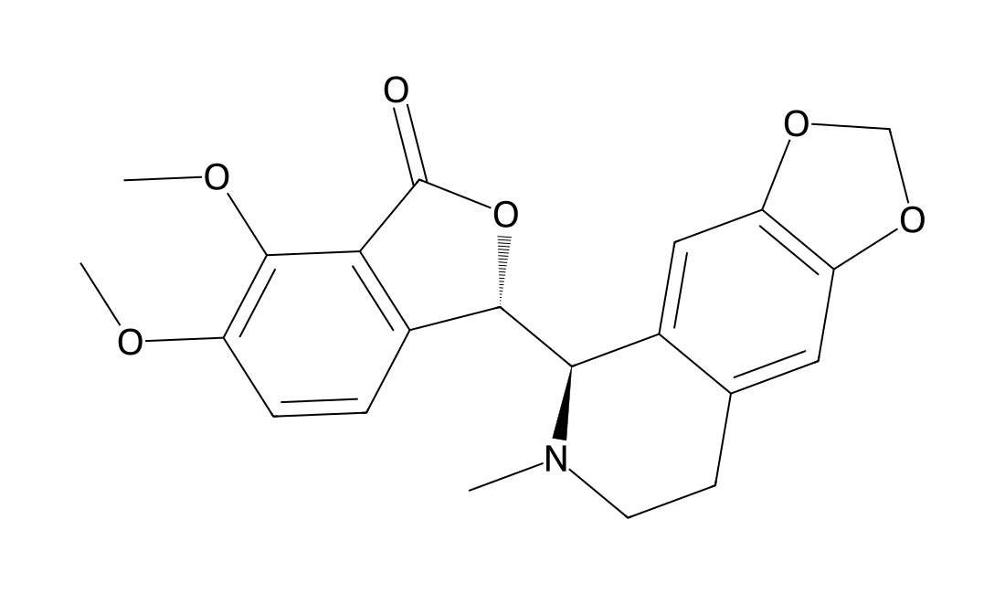
CAS 118-08-1
An alkaloid of Hydrastis canadensis used in many dietary supplements intended to enhance the immune system; inhibits PAK4 kinase activity, suppresses the proliferation and invasion of human lung adenocarcinoma cells, inhibits expression of cyclin D1/D3 and CDK2/4/6, leading to cell cycle arrest at the G1 phase; also promotes the early apoptosis of lung adenocarcinoma cells through the mitochondrial apoptosis pathway.
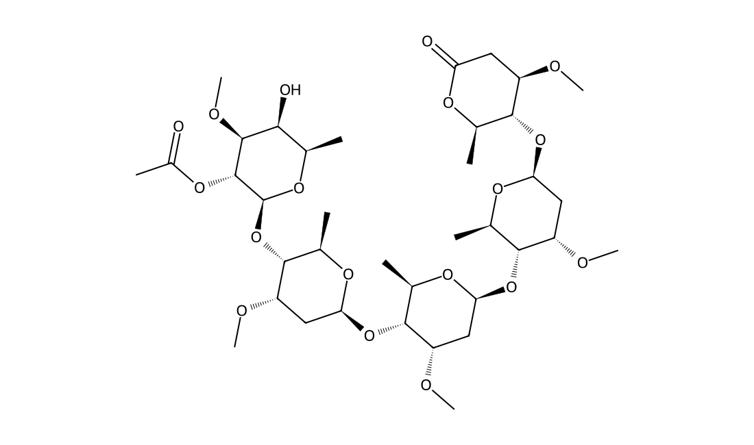
CAS 110764-09-5
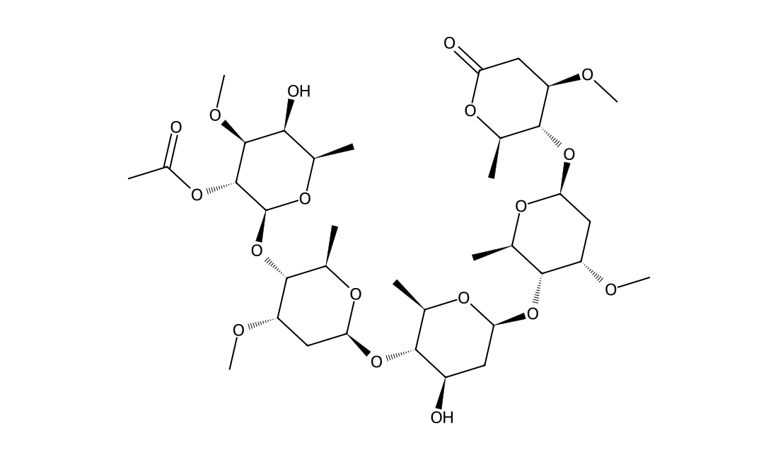
Acetyl Perisesaccharide C is an oligosaccharide, which is isolated from the root barks of Periploca sepium.
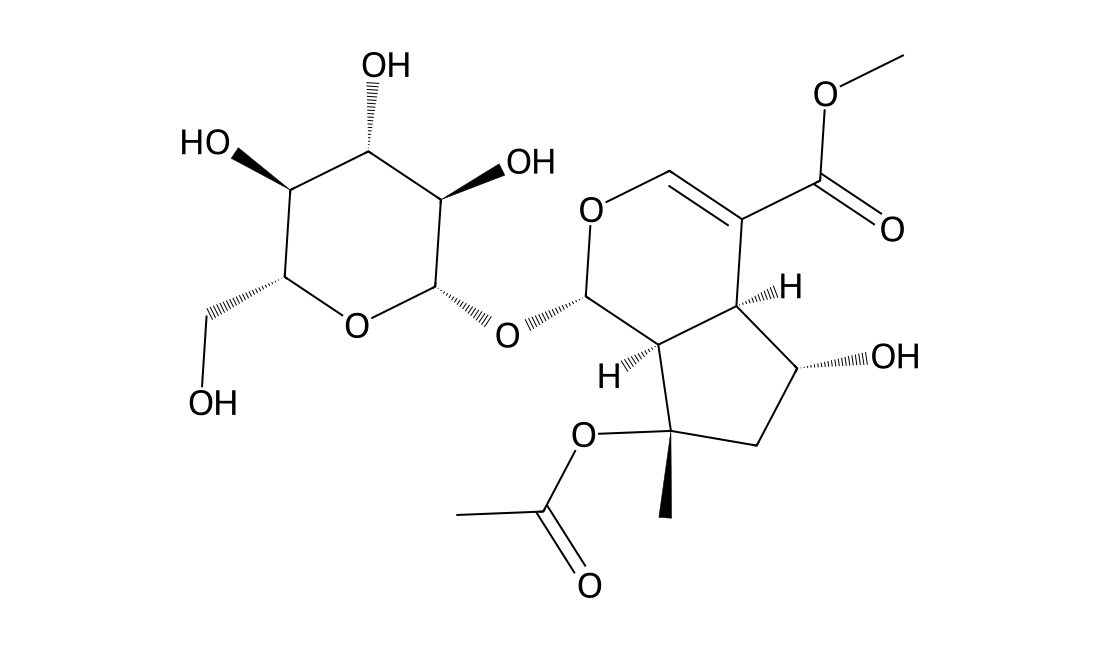
CAS 57420-46-9
Treatment of SH-SY5Y cells with ND01 blocks TNF-α-induced nuclear transcription factor κB (NF-κB) activation and decreases high-mobility group box-1 (HMGB-1) expression. . Treatment of H9c2 cells with ND01 9 μM blocks TNF-α-induced NF-κB phosphorylation by blocking High-mobility group box1 (HMGB1) expression.8-O-Acetyl shanzhiside methyl ester 40 mg/kg demonstrates significant neuroprotective effect even after delayed administration at 4 hr after I/R. 8-O-Acetyl shanzhiside methyl ester 40 mg/kg attenuates the histopathological damage, decreases brain swelling, inhibits NF-κB activation and reduces HMGB-1 expression in ischaemic brain tissue. 8-O-Acetyl shanzhiside methyl ester significantly promotes angiogenesis in the ischaemic brain and improves functional outcome after stroke. 8-O-Acetyl shanzhiside methyl ester also significantly increases vascularization compared with vehicle treatment. It increases the expression of VEGF, Ang1, phosphorylation of Tie2 and Akt VEGF. 8-O-Acetyl shanzhiside methyl ester significantly shortens capillary blood clotting time and reduces blood loss volume, but does not influence mice activated partial thromboplastin time, prothrombin time or thrombin time. It significantly prolongs euglobulin clot lysis time in hyperfibrinolysis mice.
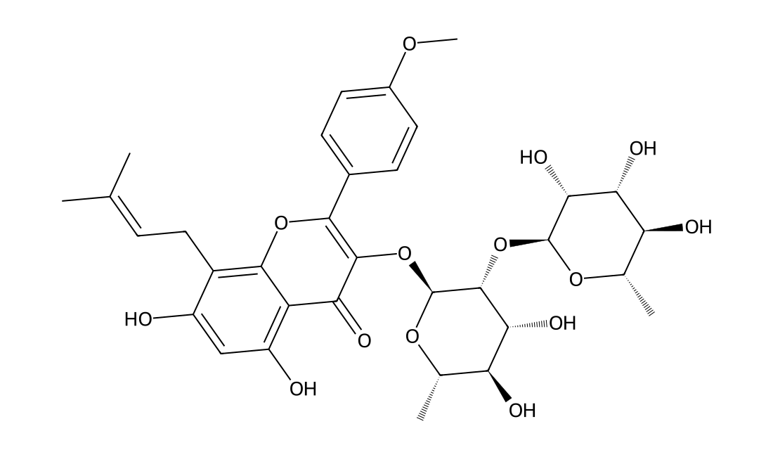
CAS 135293-13-9
2''-O-Rhamnosylicariside II is a flavonoid glycoside compound and might be beneficial for improving postmenopausal osteoporosis.