Biological Activity
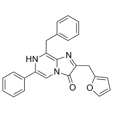
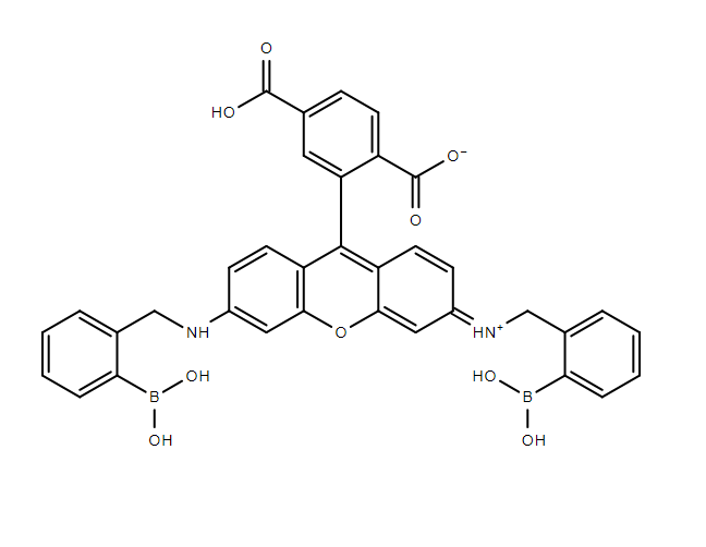
Rhobo6 is a fluorescent glycan-binding probe optimized for live-cell imaging and in vivo tracking of glycoconjugates. It exhibits pH-dependent binding (pKa ~5-7), high photostability, and minimal cytotoxicity. Compatible with mammalian cells, zebrafish, plant tissues, and mouse models.
In Vitro
Live-Cell Imaging (e.g., MCF10A)**: 1 Wash cells with serum-free PBS.2 Add 5 μM Rhobo6 in serum-free media. Incubate 15 min at 37°C. 3 Replace with fresh media. Image immediately (avoid fixation). Tissue Staining (e.g., Mouse Pancreas)** 1 Perfuse tissue with PBS. 2 Incubate with 5 μM Rhobo6 in PBS (pH 7.4) for 30 min at RT. 3 Rinse 3× with PBS. Acquire images within 2 hours.
In Vivo
1- Inject 100 μL of 1 mM Rhobo6 (in 10% DMSO/PBS) intravenously. 2-Conduct imaging 30 min post-injection (optimal distribution).
Animal Administration
1- Inject 100 μL of 1 mM Rhobo6 (in 10% DMSO/PBS) intravenously. 2-Conduct imaging 30 min post-injection (optimal distribution).
References & source
Live imaging of the extracellular matrix with a glycan-binding fluorophore-Antonio Fiore, Guoqiang Yu, Jason J. Northey,Weaver & Kayvon Pedram -Nature Methods
Published experimental findings depend on the model and assay conditions. They do not guarantee equivalent performance in every application. For laboratory research use only.