
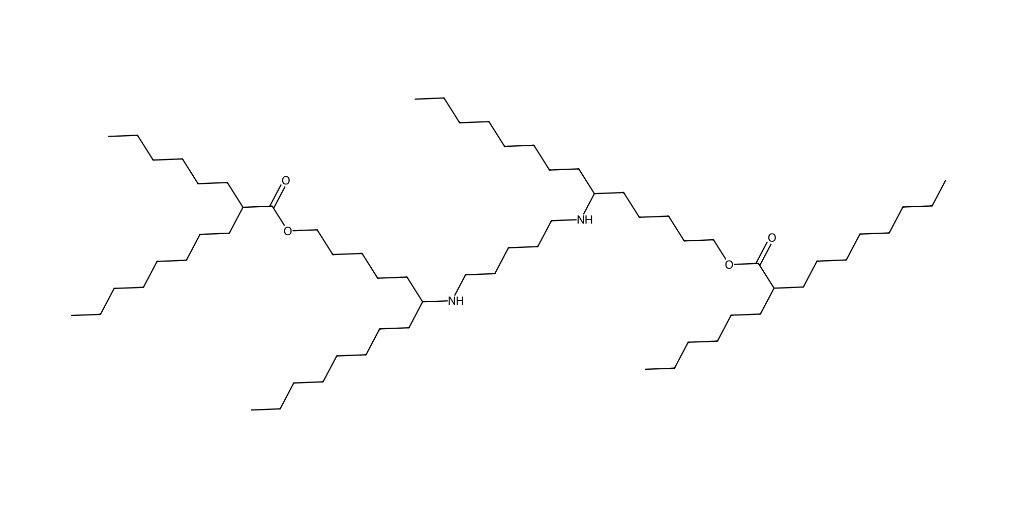
| Parameter | Value |
|---|---|
| Chemical Name | A18B8C14 |
| CAS No. | Not available |
| Synonyms | A18-B8-C14; A18 B8 C14; PILL A18B8C14 |
| Formula | C65H130N2O4 |
| M.Wt | 1003.77 |
| SMILES | CCCCCCCCC(CCCCCOC(=O)C(CCCCCC)CCCCCCCC)NCCCCCNC(CCCCCCCC)CCCCCOC(=O)C(CCCCCC)CCCCCCCC |
| Purity | >95% by ELSD-HPLC |
| Storage | Follow the supplied batch label; protect from light and moisture. Compound-specific stock-solution shelf life is not established. |
Study context and evidence coverage
The PILL study generated 275 ionizable lipids using visible-light photochemistry. A18B8C14 was selected for in vivo mRNA delivery. This guide uses the supplied 49-page Supporting Information and the publisher abstract. Exact optimized/SORT recipes and numeric main-figure endpoints await the full article.
Literature results apply to the stated formulations and test conditions; they are not batch release specifications.
Four-component formulation screen
| Study | Ionizable / helper / cholesterol / PEG-lipid | Total lipid:mRNA | Interpretation |
|---|---|---|---|
| S3a helper/PEG screen | 50:10:38.5:1.5 mol% | 20:1 w/w | DSPC + DMG-PEG2000 is one explicitly tested combination. |
| S3b ratio screen | 50:10:38.5:1.5 or 35:16:46.5:2.5 mol% | 10:1, 20:1 or 40:1 w/w | These are screening conditions. Helper/PEG identity for panel b is not specified in its caption. |
| SORT formulations | Not verified | Not verified | Abstract describes organ-selective delivery; exact recipes and effect sizes not assigned. |
Source: SI S25 Fig. S3; publisher abstract.
Helper lipids screened in S3a: DOPE, POPE, DSPC, DOPC and ESM. PEG-lipids: DMG-PEG2000, C14-PEG2000-PE and C16-PEG2000-ceramide. N/P: not reported in the verified source. The total lipid:mRNA weight ratio must not be entered as N/P.
Component molecular weights and mass conversion
The following calculation uses the tested A18B8C14/DSPC/cholesterol/DMG-PEG2000 combination at 50:10:38.5:1.5 mol%. DMG-PEG2000 uses the catalog molecular weight 2507. Values are calculated, not experimental measurements.
| Component | Molecular weight | mol% | Mass per 100 mg total lipid (mg) | Mass per 7.5 µg mRNA at 20:1 (µg) |
|---|---|---|---|---|
| A18B8C14 | 1003.77 | 50 | 65.404 | 98.105 |
| DSPC | 790.15 | 10 | 10.297 | 15.445 |
| Cholesterol | 386.65 | 38.5 | 19.399 | 29.098 |
| DMG-PEG2000 | 2507 | 1.5 | 4.901 | 7.351 |
Mass fraction = (mole fraction × molecular weight) / Σ(mole fraction × molecular weight). Total lipid for 7.5 µg mRNA at 20:1 w/w is 150 µg. These are input amounts before purification; recovery and final concentration must be measured.
Preparation and characterization
| Step / parameter | Reported method |
|---|---|
| RNA phase | 7.5 µg mRNA in 20 mM citrate, pH 3.0. |
| Lipid phase and mixing | Lipids in ethanol; aqueous:organic 3:1 v/v; rapid pipette mixing. No microfluidic flow rate is specified. |
| Post-processing | Dilute in water, process with a 10K centrifugal ultrafilter to remove residual lipids/ethanol, then dilute concentrate in DPBS. |
| Particle characterization | DLS size/zeta potential; TEM morphology; RNA fluorescence assay for encapsulation. Exact zeta/EE values are not supplied here. |
| Apparent pKa | Fluorescence assay with a four-parameter sigmoid fit; midpoint at 50% normalized signal. Exact numerical pKa is unverified. |
| N/P | Not reported; no value inferred from mass ratio. |
Source: SI S20.
Experiment-specific conditions
| Experiment | Model / cargo / dose | Route and timing |
|---|---|---|
| In vitro reporter expression | HeLa, 10,000 cells/well; Fluc mRNA 50 ng/mL | Seed 24 h before exposure; readout after 24 h. This concentration is not 50 ng/well. |
| In vivo reporter expression | 4-week male ICR CD-1 mice; Fluc mRNA 0.25 mg/kg | LNP IV; imaging after 6 h. Luciferin is administered IP for imaging. |
| Cre reporter method | Ai9 mice; Cre mRNA 0.25 mg/kg | Tail vein; tdTomato imaging and 10 µm sections after 2 days. A numerical recombination rate is not available. |
| Confocal imaging | HeLa; Cy5-mRNA LNP | 4 h exposure; LysoTracker/Cy5 Pearson correlation. |
| Hemolysis method | Rat RBCs 1% v/v; total lipid 7 µM; pH 5.5 or 7.4 | 37 °C, 1 h; 0.5% Triton positive control. No unverified hemolysis percentage assigned. |
| CBC method | 4-week male CD-1 mice; PBS, MC3 or PILL-LNP | IV; blood collection after 24 h. This timing is not automatically transferred to Fig. S9 chemistry. |
Source: SI S21–S23.
Performance, stability and mechanistic evidence
| Endpoint | Observation | Experimental context / source |
|---|---|---|
| Lead selection | The publication abstract identifies A18B8C14 as a lead and reports higher in vivo expression than the MC3 benchmark. The exact fold change and optimized/SORT compositions are not verified here. | Optimized A18B8C14 LNP versus MC3; abstract-level result only; Not available in abstract. Publisher abstract |
| Purified lipid validation | SI Fig. S5b shows stronger liver-associated luminescence for A18B8C14 than the C1/C8/C9 analogues in the displayed experiment; no image-derived fold change is assigned. | Fluc mRNA 0.25 mg/kg IV, male ICR CD-1 mice; n=4 for S5b; 6 h. SI S21, S27 Fig. S5b |
| Cellular uptake inhibition | Dynasore markedly reduces Cy5-positive uptake; other tested inhibitors show much smaller changes. The panel supports an inhibitor-sensitive uptake mechanism but does not uniquely prove a pathway. | HeLa; inhibitor pre-incubation 1 h, then Cy5-mRNA LNP exposure 1 h; 1 h LNP exposure. SI S23, S29 Fig. S7a–b |
| Endosomal colocalization | NH4Cl increases Cy5-mRNA/LysoTracker Pearson correlation; Fig. S7d marks P<0.01. This is colocalization evidence, not a measured escape percentage. | HeLa; fluorescence microscopy and Pearson correlation; 4 h general imaging protocol. SI S22, S29 Fig. S7c–d |
| Storage stability | Z-average remains approximately 150–165 nm and PDI approximately 0.06–0.17 during the displayed refrigerated series. Approximate visual readings, not tabulated values. | LNP suspension at 4 °C; caption specifies 30 days; 30-day study. SI S28 Fig. S6a |
| Freeze–thaw response | After the first cycle the plotted size is about 170 nm; subsequent cycles approach 180 nm. PDI rises to about 0.3. These observations do not establish retained mRNA expression. | −80 °C to room temperature; four cycles; approximate visual readings; 1–4 cycles. SI S20, S28 Fig. S6b |
| Dose-dependent blood chemistry | At 0.25 mg/kg all five displayed markers are marked ns versus saline. At 1 and 2 mg/kg GOT/AST and GPT/ALT increase significantly; creatinine is significantly elevated at 0.5, 1 and 2 mg/kg. ALP is marked ns at all doses. | Fluc-mRNA A18B8C14 LNP IV; 0.25, 0.5, 1 and 2 mg/kg; n=4. Blood-chemistry sampling time is not specified in Fig. S9.; Not specified for Fig. S9. SI S31 Fig. S9 |
Dose-dependent interpretation
Fig. S9 does not support an unrestricted “nontoxic” claim. Although the 0.25 mg/kg group is marked nonsignificant versus saline for the displayed markers, higher doses increase transaminases and creatinine. The experiment has n=4, and nonsignificance is not equivalence or clinical safety.
Structure, synthesis and analytical identity
The two secondary amines are connected by a five-carbon linker. Each arm contains an α-branched amine and a 2-hexyldecanoate ester. The structure and formula C65H130N2O4 are explicitly shown in S19. Four stereocenters are unassigned.
A18 (1,5-diaminopentane), B8 and C14 were reacted under 427 nm irradiation for 4 h at room temperature for this compound. The general procedure uses HFIP and catechol, followed by preparative chromatography. Formic acid/ammonium formate in the mobile phase does not establish an isolated formate salt.
| Analytical record | Reported value / interpretation |
|---|---|
| Average molecular weight | 1003.77 (SI S19); structure-based calculation agrees within rounding. |
| HRMS [M+H]+ | Calculated 1004.0103; found 1004.0117. |
| HRMS [M+2H]2+ | Calculated 502.5088; found 502.5067. |
| Research material HPLC | 96.00% stated in S19; S48 UV 210 nm peak table gives 96.005%. This is not a DC Chemicals batch result or ELSD-HPLC measurement. |
| NMR / mass spectra | 1H/13C NMR and HPLC S48; LRMS/HRMS S49. |
| Simulation | 50 lipids, Martini 3 / GROMACS 2024, 1 µs. PILL +2 at modeled pH 5.5 is a simulation assumption, not a measured pKa or isolated salt assignment. |
Source: SI S12, S19, S24, S48–S49.
References
Chun S, Nam H, Lee D, Lee Y, Park CH, Lim C, Kim D, Park JS, Kim KP, Choi HS, Koo H. Photochemistry-Based Development of Ionizable Lipid Library for mRNA-Lipid Nanoparticle Delivery In Vivo. Journal of the American Chemical Society. 2026;148(39):42494–42507. doi:10.1021/jacs.6c13674. Article and Supporting Information.