Biological Activity
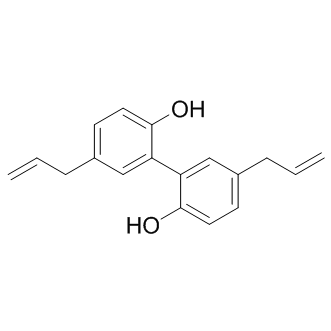
Magnolol, a natural lignan isolated from the stem bark of Magnolia officinalis, is a dual agonist of both RXRα and PPARγ, with EC50 values of 10.4 µM and 17.7 µM, respectively.
In Vitro
Magnolol is a dual agonist of both RXRα and PPARγ, with EC50 values of 10.4 µM and 17.7 µM, respectively. Magnolol (26.2-80 µM) binds to RXRαLBD and PPARγLBD in a dose dependent manner, with Kd values of 45.7 µM and 1.67 µM, respectively. Magnolol (1-20 µM) induces the transcription of PPRE in a dose-dependent manner, but shows no activity on RXRE transcription[1]. Magnolol (1, 3, 10 µM) enhances adipocyte differentiation of both 3T3-L1 pre-adipocystes and C3H10T1/2 pluripotent stem cells in the presence of insulin. Magnolol (10 μM) upregulates mRNA expression of marker genes for adipocyte differentiation. Magnolol (1, 10 μM) shows an increase in basal and insulin-stimulated glucose uptake in differentiated 3T3-L1 adipocytes[2].
In Vivo
Magnolol (5-15 mg/kg, p.o.) significantly attenuates the phenotypic severity of dextran sulfate sodium (DSS)-induced colitis in mice. Magnolol (10, 15 mg/kg, p.o.) attenuates histopathological changes and myeloperoxidase activity in the colon of DSS-treated mice, decreases DSS-induced high levels of proinflammatory cytokines TNF-α, IL-1β and IL-6 in the colonic tissues. Magnolol (10 mg/kg, p.o.) also reverses abnormality of serum metabolome, and regulates tryptophan metabolic pathway in mice[3].
Target
RXRα:10.4 μM (EC50)
PPARγ:17.7 μM (EC50)
Kinase Assay
Binding affinities of magnolol towards purified RXRαLBD and PPARγLBD are analyzed using Biacore 3000 instrument. Proteins are covalently immobilized to CM5 chip using a standard amine-coupling procedure in 10 mM sodium acetate buffer (pH 4.2). The chip is equilibrated with a continuous flow of running buffer (10 mM HEPES, pH 7.4, 150 mM NaCl, 3 mM EDTA, 0.005% (v/v) surfactant P20) for 2 hours. Subsequently, magnolol in a gradient of concentrations are injected into the channels at a flow rate of 20 µL/min for 60 seconds, followed by disassociation for 120 seconds. For the coactivator SRC1 recruitment assays, biotin-labelled SRC1 is immobilized to SA chip. Different concentrations of Magnolol are incubated with 5 µM RXRαLBD or PPARγLBD for 1 hour, and then injected to the channel at a flow rate of 20 µL/min for 60 s, followed by disassociation for 120 s[1].
Cell Assay
For differentiation of 3T3-L1 pre-adipocytes, at 2 days after confluence (defined as day 0), cells are incubated in differentiation medium containing 0.5 mM IBMX, 10 μg/mL insulin and 0.25 μM DEX in DMEM containing 10% fetal bovine serum (FBS). After 2 days, the cell culture medium is changed to DMEM containing 10 μg/mL insulin and 10% FBS. The medium is replaced again with fresh DMEM containing 10% FBS after 2 days. Adipocytes are used 6-8 days after the initiation of differentiation. In adipogenesis studies, 3T3-L1 pre-adipocytes and C3H10T1/2 pluripotent stem cells grown in DMEM supplemented with 10% bovine calf serum (day 0) are treated with insulin (1 μg/mL) with/without Magnolol in 10% FBS contained DMEM at the indicated concentration for 9 days. Fresh medium containing insulin (1 μg/mL) and 10% FBS with/without magnolol is replenished every 3 days[2].
Animal Administration
Experimental colitis mice model is induced by routine administration of dextran sulfate sodium (DSS) solution dissolved in drinking distilled water at a concentration of 2.0% (w/v) ad libitum for 5 consecutive days. Distilled water is given to mice in the normal group for the same period. The body weight of each mice is recorded daily in the morning (9:00 a.m.). On day 6, the mice with significant body weight loss, diarrhea, and gross bleeding are considered as experimental candidates of colitis. All the mice with comparable disease index are then randomly divided into 5 groups (n = 8/group): (1) DSS model group, intragastric administrated with saline; (2) positive control group, intraperitoneal injected with infliximab (5 mg/kg); (3) low dose treatment group, intragastric administrated with Magnolol (5 mg/kg); (4) medium dose treatment group, intragastric administrated with Magnolol (10 mg/kg); (5) high dose treatment group, intragastric administrated with Magnolol (15 mg/kg). The mice in control group receives drinking water without DSS throughout the entire experimental period and intragastric administrated with saline[3].
References & source
[1]. Zhang H, et al. Molecular determinants of magnolol targeting both RXRα and PPARγ. PLoS One. 2011;6(11):e28253.
[2]. Choi SS, et al. Magnolol enhances adipocyte differentiation and glucose uptake in 3T3-L1 cells. Life Sci. 2009 Jun 19;84(25-26):908-14.
[3]. Zhao L, et al. Magnolol, a Natural Polyphenol, Attenuates Dextran Sulfate Sodium-Induced Colitis in Mice. Molecules. 2017 Jul 20;22(7). pii: E1218.
Published experimental findings depend on the model and assay conditions. They do not guarantee equivalent performance in every application. For laboratory research use only.