Formulation & In Vivo Data
Lipid P3B · DC60934 · Updated 29 October 2026
Formulation & In Vivo Data
A reference lipid for research on intrathecal mRNA delivery and functional gene editing in the central nervous system.
Values shown are literature-reported results for the cited formulation and study conditions. They do not guarantee performance of the supplied product in other formulations or experiments.
Formulation and particle properties
DOE-03 was selected in the reported formulation comparison. The record below separates consistent findings from unresolved composition labels.
| Parameter | Literature record | Source / status |
|---|---|---|
| Delivery route | Intrathecal; mouse CNS research | Figures 2–5 / Reported |
| Study cargos | Reporter mRNA, Cas9 mRNA with guide RNA, and adenine base-editor mRNA with guide RNA | Figures 2–4 / Reported |
| Ionizable lipid:mRNA | 10:1 by mass | Formulation optimization section / Reported; not an N/P value |
| N/P ratio | Unresolved | Figure 2a axis labeling requires reconciliation with the mass-ratio description |
| Hydrodynamic diameter / PDI | 110 nm / 0.108 | Formulation optimization section / Reported |
| LNP apparent pKa | 6.51 | Main text citing Figure S16; supporting assay details not independently verified |
| Particle morphology | Approximately spherical particles | Figure 2f / Reported |
| Encapsulation efficiency / zeta potential | No verified numerical value entered | Supporting information review pending |
Component identity, molecular weight and reported molar fraction
| Component | MW | Optimized formulation | DC Chemicals product |
|---|---|---|---|
| P3B | 645.07 | 60 mol% reported | DC60934 |
| DOPE | 744.0478 | 30 mol% reported | DC33634 |
| Cholesterol | 386.65 | Not assigned while PEG fraction is unresolved | DCD-041 |
| PEG-lipid | Study identity pending; catalog DMG-PEG2000: 2507 | 0.5 mol% in text; 1.5% in Figure 2e | DMG-PEG2000 · DC40169 |
Composition review: the main text and Figure 2e disagree on PEG content. The paper also uses C14-PEG and DMG-PEG labels. A definitive four-component molar-to-mass conversion is therefore withheld. Catalog molecular weights above are reference values, not confirmation of the paper's PEG reagent identity.
Functional CNS delivery
| Model and readout | Reported finding | Context |
|---|---|---|
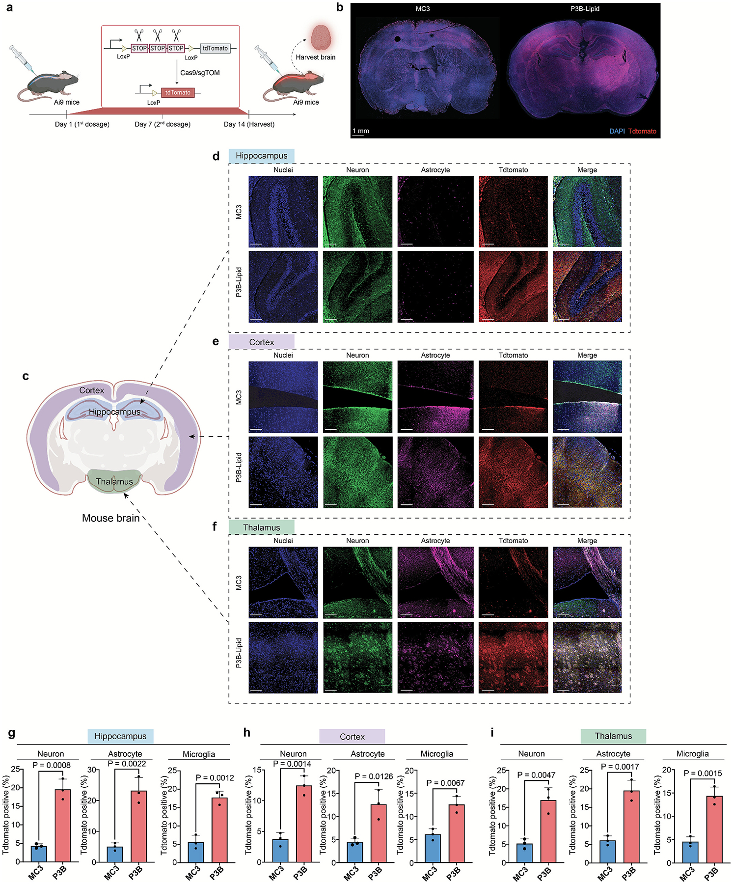
| Ai9 reporter mice; Cas9-mediated reporter activation | Higher tdTomato-positive cell fractions than the MC3 comparator in the examined CNS regions | Figure 3; intrathecal; 0.05 mg/kg mRNA; repeat administration; day-14 assessment; n=3 biological replicates |
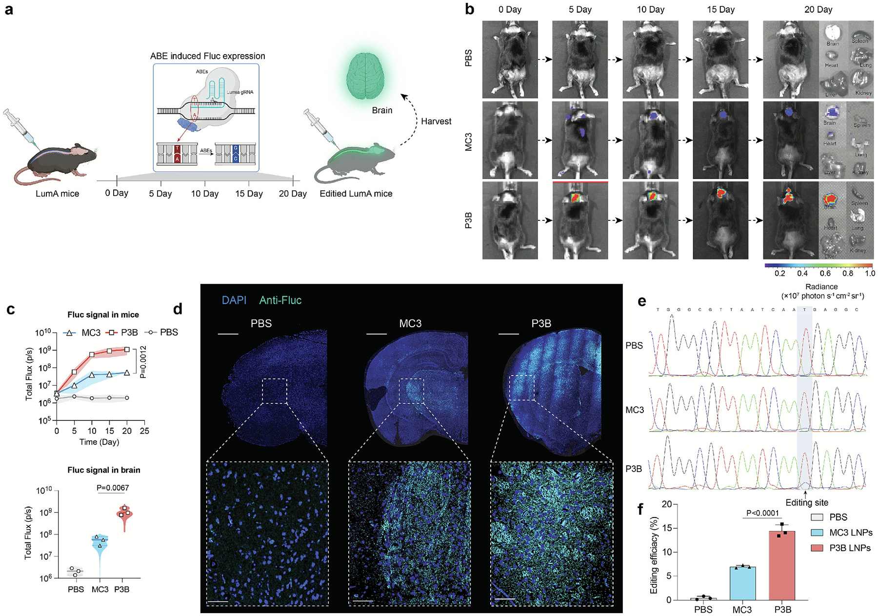
| LumA reporter mice; target-site base editing | P3B: 14.8%; MC3: 6.9% | Figure 4e–f; intrathecal; 0.05 mg/kg mRNA; day-20 assessment; Sanger/ EditR; n=3 |
| Editing-rate comparison | Approximately 2.14-fold (14.8 ÷ 6.9) | Calculated from the reported editing percentages; not the luminescence ratio |
| Reporter signal persistence | Signal observed from day 5 through day 20 | Figure 4b–c; reflects the reporter-editing experiment, not 20-day persistence of intact LNPs |
These are reporter-model findings. They establish functional delivery under the studied conditions; intravenous blood–brain barrier crossing and disease-treatment efficacy were not demonstrated by these experiments.
Regional percentages are not transcribed here because some cortex/thalamus values in the text do not match the Figure 3 panel labels. The original panels are shown with their published labels.
Acute tolerability
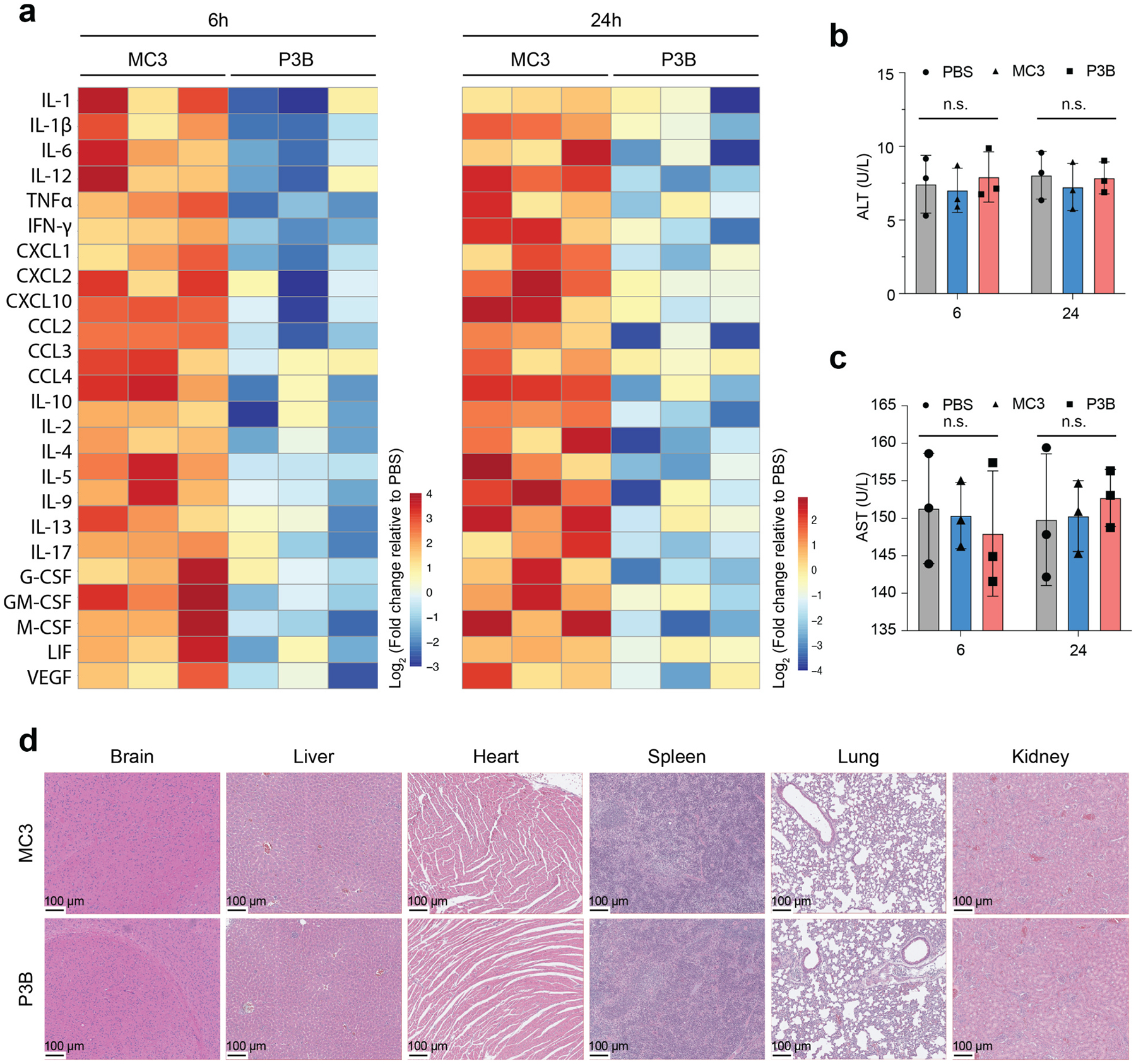
The study reported a weaker inflammatory-marker response with P3B than with MC3. Plasma ALT and AST did not show significant between-group changes at the measured 6- and 24-hour endpoints. Histology at 6 hours did not show obvious tissue injury in the examined organs (Figure 5; n=3).
This is short-term preclinical evidence. The authors identify the 24-hour safety observation window as a limitation; long-term and repeated-dose safety remain to be established. The text and Figure 5 caption also differ in the stated sample compartment for inflammatory-marker profiling, so no tissue-specific cytokine claim is assigned here.
Reference and evidence basis
Dong S, Healy L, Gong F, et al. Biodegradable lipid nanoparticles for genome editing in the brain via intrathecal administration. Materials Today. 2026;92:140–150. doi:10.1016/j.mattod.2025.11.032.
Read the full paper · PubMed record
Structure identity: Figure 1f, P3B / H8TA2TB5. Formula and molecular weight were calculated from that structure; the newly formed stereocenter is unspecified. Main text and Figures 1–5 were reviewed. Supplementary files were not independently verified in this release.